
Figure 2.
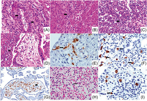
Multi‐organic fetal and placental inflammation. Histological sections with haematoxylin/eosin and immunohistochemistry from the postmortem study of fetus A and placenta showing severe inflammation. (A and B) Arrows point to inflammatory cells (macrophages and neutrophils) in the intima, media and adventitia of the arterial vessels of the heart as well as between the fascicles of the cardiac muscle cells, with several CD68‐positive macrophages (E, brown stain) (×20). (C) Fetal lung shows mild interstitial hypercellularity with inflammatory cells which express CD68 (F, arrows, brown stain) (×20). (D) Chorionic villi with active chronic intervillositis and abundant perivillous neutrophils (arrow) (×10). (G) Section of the placenta exhibiting severe inflammation with Hofbauer cells in the villous stroma strongly positive for CD163 cells (arrow, brown stain) (×10). (H) Striated muscle with inflammatory infiltrates of mononuclear cells and neutrophils, as well as edema, apoptosis, and diffuse myocyte atrophy (arrow, ×40). (I) Immunohistochemical staining for CD68 shows interstitial macrophages infiltrate in the kidney (brown stain) (×40)