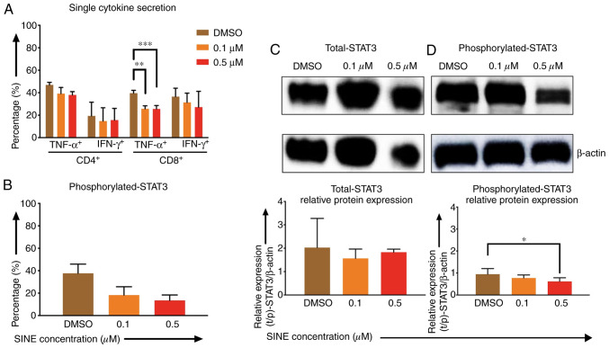
Figure 4.
Effects of SINEs when eltanexor, CAR T cells and Daudi cells were cultivated simultaneously. Daudi cells were cultivated simultaneously with CAR T cells and eltanexor (0.1 µM: Orange, 0.5 µM: Red) or with DMSO as control (brown) for 6 h (A, assessed via flow cytometry analysis) or 4 h (B, assessed via flow cytometry analysis, C and D via western blot analysis). Cytokine release (IFN-γ and TNF-α) of CD4- and CD8-positive CAR T cells was assessed and decreased cytokine secretion after stimulation of CAR T cells with Daudi cells compared to the DMSO control was observed. (A) Phosphorylated-STAT3 level of CAR T cells was evaluated. Decreased phosphorylated-STAT3 of CAR T cells cultured with eltanexor was observed when compared to the DMSO control. (B) Evaluation of total STAT3 and phosphorylated-STAT3 protein levels of CAR T cells was performed after protein isolation via western blot analysis on STAT3 of CAR T cells; β-actin was used as the internal reference. Raw total STAT3 protein level (C, upper panel), relative STAT3 expression (C, lower panel), raw phosphorylated-STAT3 protein level (D, upper panel) and relative phosphorylated-STAT3 expression (D, lower panel) were assessed. The protein levels of phosphorylated-STAT3 compared to total STAT3 were decreased. Experiments were performed in triplicate. Mean values were calculated for each group; results are presented as mean ± standard deviation (SD). *P<0.05, **P<0.005, ***P<0.0005 indicates statistical significance.

