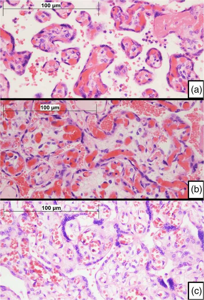
Figure 1.

Microscopic features of placentas. (a) Normal term villi. Figure shows slender villi with a high proportion of capillary vessels forming vasculosyncytial membranes, with little intervening stroma. A thin layer of intervening syncytiotrophoblast can be seen. Although congestive, villi do not fulfill criteria for diagnosis of villous chorangiosis (H&E ×400). (b) Villous chorangiosis. Villi show an increased number of capillary cross‐sectional profiles per villus (per definition, at least 10 capillaries per villus are required to fulfill minimal criteria for villous chorangiosis) (H&E ×400). (c) Delayed villous maturation. Terminal villi show features that resemble immature intermediate villi. Villi tend to be larger, with an increased amount of stroma, which may be edematous. There is usually a thick layer of syncytiotrophoblast with scattered persistent cytotrophoblast cells. Villous capillaries are nonperipherical and do not to merge with the overlying syncytiotrophoblast, failing to form vasculosyncytial membranes. Delayed villous maturation may be seen in association with villous chorangiosis. Compare villous features with A (H&E ×400)
