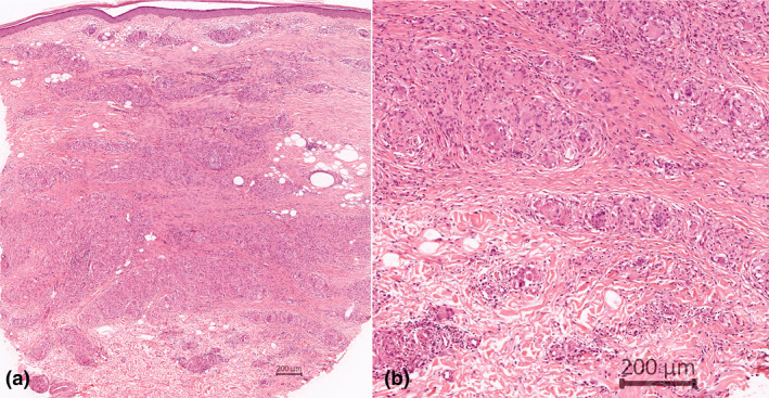
Figure 2.

(a) Panoramic view of punch biopsy shows granulomatous inflammation in the entire dermis, especially in the lower half (HE [haematoxylin–eosin]); (b) well‐delimited granulomas formed by histiocytes and many giant cells. Few lymphocytes participate (HE).
