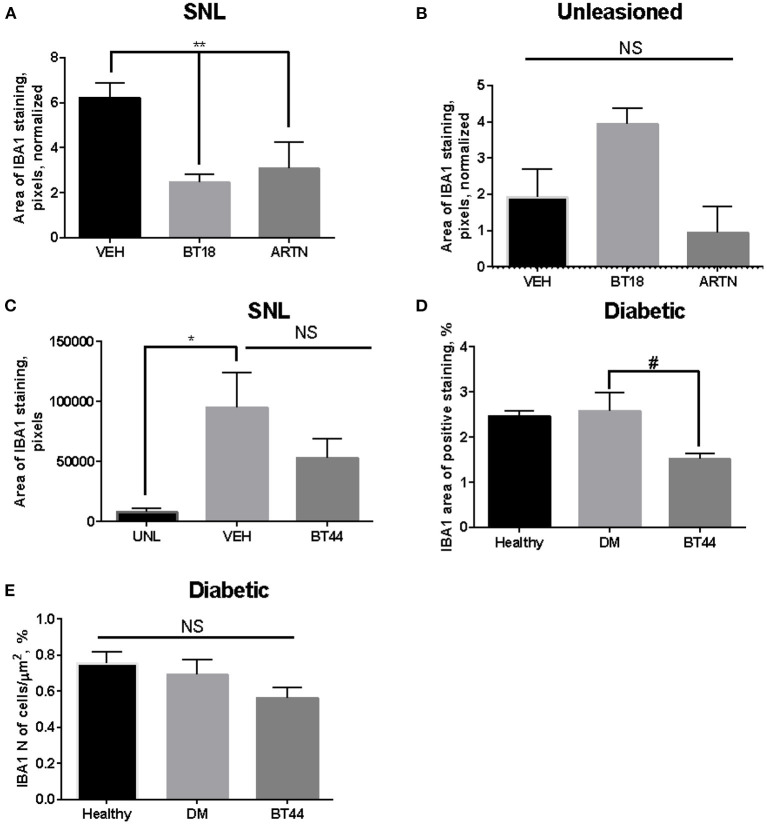
Figure 2.
Expression of IBA-1 in dorsal root ganglia (DRG) of animals with surgery (spinal nerve ligation, A–C) or diabetes-induced neuropathy (D,E). (A) Expression of IBA1 (area) in DRGs which spinal nerve was ligated upon the treatment with vehicle (VEH), BT18 (the first generation RET agonist, subcutaneously at the dose 25 mg/kg every other day for 12 days, started 1 h after the lesion), and ARTN (at the dose 0, 5 mg/kg every other day for 12 days, started 1 h after the lesion), n = 3 per group. (B) Expression of IBA1 in DRGs on the unligated side of the body (healthy), the treatments and doses are the same as in (A), n = 3 per group. (C) Expression of IBA1 (area) in DRGs which spinal nerve was ligated upon the treatment with vehicle (VEH) or BT44 (the second generation RET agonist, subcutaneously at the dose 12.5 mg/kg every other day for 12 days, started 2 days after the lesion) or on unleasoned side (UNL), n = 3–4 per group. (D) Expression of IBA1 (area) in DRGs of healthy animals (healthy) or animals with diabetes-induced neuropathy treated subcutaneously with vehicle (DM) or BT44 (at the dose 5 mg/kg every other day for 3 weeks started on the day of lesion), n = 4–5 per group. (E) As in (D), but the number of cells is calculated, n = 5 per group, *p < 0.05, **p < 0.01 by ANOVA and post-hoc test, #p < 0.05 by post-hoc test (ANOVA p = 0.059).

