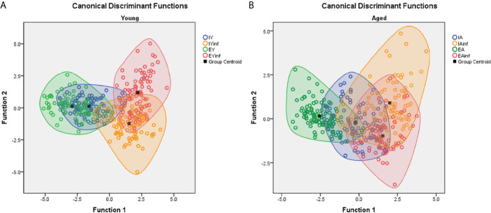
Figure 7.
Discriminant function analysis to illustrate age and environmental influences on microglial morphological response in a mouse model of sublethal encephalitis induced with Piry arbovirus. Note that in young mice (A) there is a distinct distribution of microglia of infected (red and orange color open circles) in the right superior and inferior quadrants respectively, while in controls, the green and blue color open circles are in the central area of left quadrant. In contrast, the microglia of aged mice (B), show larger areas of intersection between infected and control mice, indicating smaller influence of environment on microglia morphological response. Orange and red circles represent infected mice; Green and blue circles indicate control mice. IY, young mice raised in impoverished environment; EY, young mice raised in enriched environment; IA, aged mice raised in impoverished environment; EA, aged mice raised in enriched environment; IYinf, infected young mice raised in impoverished environment; EYinf, infected young mice raised in enriched environment; IAinf, infected aged mice raised in impoverished environment; EAinf, infected aged mice raised in enriched environment. Square black dot = ellipsoid center of each experimental group.

