

Abstract
COVID‐19 leads to severe respiratory problems, but also to long‐COVID syndrome associated primarily with cognitive dysfunction and fatigue. Long‐COVID syndrome symptoms, especially brain fog, are similar to those experienced by patients undertaking or following chemotherapy for cancer (chemofog or chemobrain), as well in patients with myalgic encephalomyelitis/chronic fatigue syndrome (ME/CFS) or mast cell activation syndrome (MCAS). The pathogenesis of brain fog in these illnesses is presently unknown but may involve neuroinflammation via mast cells stimulated by pathogenic and stress stimuli to release mediators that activate microglia and lead to inflammation in the hypothalamus. These processes could be mitigated by phytosomal formulation (in olive pomace oil) of the natural flavonoid luteolin.
Keywords: brain fog, chemotherapy, coronavirus, COVID‐19, cytokines, fatigue, inflammation, mast cells, microglia
Abbreviations
- AD
Alzheimer's disease
- ACE2
angiotensin converting enzyme 2
- BBB
blood–brain barrier
- CNS
central nervous system
- CRH
corticotropin‐releasing hormone
- DAMPs
damage‐associated molecular patterns
- HPA
hypothalamic–pituitary–adrenal
- MCAS
mast cell activation syndrome
- MCI
mild cognitive impairment
- mtDNA
mitochondrial DNA
- ME/CFS
myalgic encephalomyelitis/chronic fatigue syndrome
- PAMPs
pathogen‐associated molecular patterns
- PAF
platelet activating factor
- SP
substance P
- SM
systemic mastocytosis
- TLCOVID‐19
toll‐like receptor
1. INTRODUCTION
Infection with the recent coronavirus (severe acute respiratory syndrome [SARS]‐CoV‐2) leads to COVID‐19, the severity of which derives from the host's inflammatory response that involves release of a storm of pro‐inflammatory cytokines, 1 , 2 , 3 , 4 , 5 , 6 , 7 especially interleukin‐6 (IL‐6), 8 , 9 , 10 , 11 but also IL‐1. 12 , 13
Even though symptoms associated with SARS‐CoV‐2 infection in children are mild, a number of recent publications reported a multisystem inflammatory syndrome (MIA‐C) in older children 14 , 15 , 16 and adolescents, 17 often presenting with symptoms reminiscent of Kawasaki disease. 16 Symptoms in MIA‐C typically occur 4–6 weeks after infection and the disease is characterized by elevated markers of inflammation 18 and the presence of multiple autoantibodies. 18 A similar disease in adults, named multisystem inflammatory syndrome (MIA‐A) has been recognized by the Center for Disease Control (CDC, USA) (https://www.cdc.gov/mis-c/). In fact, autoimmune and inflammatory diseases are now increasingly identified following COVID‐19. 19 The etiology of MIA remains unknown.
Cytokine storms have also been implicated in a variety of “mystery” diseases. 20 One such disease affects COVID‐19 survivors and is associated with severe fatigue and neuropsychiatric symptoms (https://www.health.harvard.edu/blog/the‐tragedy‐of‐the‐post‐covid‐long‐haulers‐2020101521173), especially impairment in cognitive functions known as “brain fog” (https://www.nytimes.com/2020/10/11/health/covid‐survivors.html). Such patients have been called “long‐haulers” (https://directorsblog.nih.gov/tag/post-covid-syndrome/) and the illness has been termed “long‐COVID syndrome” (https://directorsblog.nih.gov/2021/01/19/trying‐to‐make‐sense‐of‐long‐covid‐syndrome/). In fact, the National Institutes of Health (NIH, USA) recently devoted a 2‐day conference on the epidemiology and pathophysiology of this illness (https://www.niaid.nih.gov/news‐events/workshop‐post‐acute‐sequelae‐covid‐19). Other names used for this illness include “chronic COVID syndrome,” “post‐COVID syndrome,” or “long haulers COVID syndrome.” 21
In addition to the severe respiratory and inflammatory problems discussed above, infection with SARS‐CoV‐2 can also contribute to neurological 22 , 23 , 24 , 25 and mental 26 , 27 , 28 , 29 , 30 disorders. For this reason, NIH held an 1‐day workshop on the effect of COVID‐19 on the central nervous system (CNS) (https://www.ncbi.nlm.nih.gov/search/research-news/11277/ ) and recently launched a database to track neurological symptoms associated with COVID‐19 (https://www.nih.gov/news‐events/news‐releases/nih‐launches‐database‐track‐neurological‐symptoms‐associated‐covid‐19). The importance of the effects of COVID‐19 on the brain is highlighted by the blog recently posted by the NIH Director on this subject (https://directorsblog.nih.gov/2021/01/14/taking-a-closer-look-at-the-effects-of-covid-19-on-the-brain/).
However, few scientific publication has so far discussed long‐COVID syndrome (https://www.nytimes.com/2021/01/21/magazine/covid-aftereffects.html) such as the one that reported the presence of persistent fatigue apparently independent of the severity of the initial symptoms. 31 Symptoms experienced by long‐COVID syndrome patients (Table 1) are very similar 32 to those present in patients with myalgic encephalomyelitis/chronic fatigue syndrome (ME/CFS), 33 , 34 mast cell activation syndrome (MCAS), 35 , 36 or systemic mastocytosis (SM) 37 in whom the unique tissue immune cells, mast cells, are stimulated by environmental, pathogenic, and stress stimuli. Moreover, IL‐6 has not only been implicated in COVID‐19 8 , 12 but was also elevated in ME/CFS 38 and SM. 39 , 40 , 41 To make matters worse, IL‐6 promotes an increase in number of mast cells. 42
TABLE 1.
Symptoms present in long‐COVID syndrome
|
|
|
|
|
|
|
|
|
|
|
|
|
|
|
|
|
|
|
Note: The symptoms listed, especially those bolded, are experienced, by many long‐COVID syndrome patients and also patients undergoing or after having been administered chemotherapy.
2. CHEMOTHERAPY
Patients undergoing chemotherapy are susceptible to infection with COVID‐19. 43
Moreover, more than 50% of patients on or following chemotherapy develop symptoms similar to those described above for long‐COVID syndrome (Table 1), especially cognitive dysfunction, 44 , 45 , 46 , 47 a condition that has been termed “chemofog” 48 , 49 or “chemobrain,” 50 , 51 , 52 , 53 , 54 and has been associated with distinct neuroimaging findings. 55 , 56 A number of drugs have been implicated in “chemobrain” (Table 2) most notably doxorubicin, 57 , 58 , 59 methotrexate, 60 , 61 lenalidomide, 62 rituximab, 62 and trastuzamab. 63
TABLE 2.
Chemotherapeutic agents implicated in chemofog
|
|
|
|
|
|
|
|
|
|
|
Note: The drugs listed, especially those bolded, have been reported to induce “chemofog” or “chemobrain”.
There have been intense efforts to understand the biochemical 64 or cellular 44 , 65 , 66 mechanisms responsible for chemobrain. These have included disrupted neurogenesis, 67 aberrant myelination, 68 , 69 interference with prefrontal activity, 70 but most importantly neuroinflammation 66 with cytokine dysregulation. 69 , 71
3. INFLAMMATION OF THE BRAIN
Microglia have important functions in the CNS, 72 especially with respect to neuroinflammation 72 , 73 , 74 and neurodegenerative 75 , 76 , 77 diseases. Microglia express Toll‐like receptors (TLRs), 78 activated by damage‐associated molecular patterns (DAMPs) and were recently implicated in COVID‐19. 79 , 80 COVID‐19 can also affect the hypothalamic–pituitary–adrenal (HPA) axis, 81 which is typically activated by stress and can further affect the emotional state of individuals affected by COVID‐19. 82 , 83 Microglia also express receptors for corticotropin‐releasing hormone (CRH) 84 and could be further activated by stress, especially associated with COVID‐19. 85
Microglia interact with the unique immune cells, mast cells, in the brain 86 leading to their activation 87 and neuroinflammation. 88 Activation of mast cells 89 , 90 and microglia 91 in the hypothalamus 33 could lead to cognitive dysfunction 92 commonly also seen in patients with MCAS 93 , 94 (Figure 1). Psychological stress has pro‐inflammatory effects 82 , 95 via stimulation of mast cells, 83 especially by CRH 96 leading to increased vascular permeability. 83 This process also leads to disruption of the blood–brain barrier (BBB), 97 , 98 via release of IL‐6 99 and CRH, 100 further exacerbating brain inflammation by permitting the entry into the brain of more viral particles, cytokines, or other toxic substances (Table 1). A recent NIH study reported blood vessel damage and inflammation, but no infection, in brains of patients with COVID‐19. 101 (https://www.nih.gov/news‐events/news‐releases/nih‐study‐uncovers‐blood‐vessel‐damage‐inflammation‐covid‐19‐patients‐brains‐noinfection#:~:text=In%20an%20in%2Ddepth%20study,shortly%20after%20contracting%20the%20disease.) These findings may explain the recent comprehensive reports of significantly increased neurologic 102 and psychiatric 103 disorders in COVID‐19 patients, as well as in long‐COVID syndrome patients. 104
FIGURE 1.
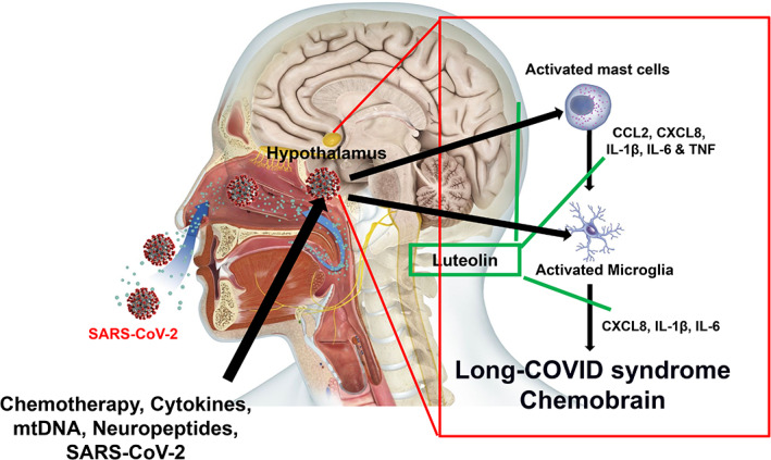
Diagrammatic representation of how SARS‐CoV‐2 could stimulate mast cells and microglia in the hypothalamus, inhibited by luteolin. SARS‐CoV‐2 could enter the brain via the olfactory nerve tract reaching the hypothalamus where it could activate brain mast cells and microglia to release pro‐inflammatory molecules, thus contributing to brain inflammation and brain fog. The effect of SARS‐CoV‐2 could be exaggerated by chemotherapy, as well as cytokines and mtDNA, or neuropeptides released under stress (thunderbolt). These processed could contribute to the pathogenesis and symptoms of long‐COVID syndrome and “chomobrain” and could be prevented by the flavonoid luteolin
Mast cells are ubiquitous in the body 37 and are critical for allergic diseases, 105 but also inflammation. 106 Mast cells are also present in the brain, especially in the median eminence of the hypothalamus, where they are located perivascularly close to nerve endings positive for CRH. 107 Mast cells are also triggered by viruses 108 including SARS‐CoV‐2. 109 , 110 A recent publication using normal oral cavity mucosa reported no gene expression of the SARS‐CoV‐2 receptor, angiotensin converting enzyme 2 (ACE2) in mast cells. 111 However, mast cells are “plastic” and their surface receptors can be induced by a variety of conditions. For instance, we reported that the neuropeptides neurotensin 112 and substance P (SP) 113 can induce CRHR‐1. Moreover, SP can induce the ST2 receptor for IL‐33. 114 In fact, ACE2 gene expression was recently shown to be induced by interferon, 115 and mast cells can elicit strong pro‐inflammatory and type I interferon responses in response to viruses, 116 implying an autocrine action on ACE2 expression. Of course, it remains to be seen to what extent pulmonary and/or brain mast cells from deceased COVID‐19 patients express ACE2.
Following stimulation, mast cells release pro‐inflammatory mediators 117 such as histamine, tryptase, chemokines (e.g., CCL2, CCXL8) 118 and cytokines (IL‐6, 119 IL‐1β , 120 and tumor necrosis factor [TNF] 114 ), especially when primed by IL‐33. 121 , 122 Histamine can stimulate macrophages to release IL‐1, 123 which stimulates mast cells to release IL‐6. 119 Mast cells can also secrete mitochondrial DNA (mtDNA), 124 which was recently reported to be increased in the serum of COVID‐19 patients and correlated with disease severity. 125 Extracellular mtDNA serves as an alarmin and stimulates pro‐inflammatory mediator secretion from immune cells. 126 , 127 Moreover, mast cells synthesize and release platelet activating factor (PAF), which has been implicated in the inflammation 128 and microthromboses 129 characterizing COVID‐19.
4. TREATMENT APPROACHES
Unfortunately, there are no clinically effective interventions for long‐COVID syndrome 1 , 130 or brain fog associated with either chemobrain, ME/CFS, 131 or MCAS. 36 It is also hard to decide whether it would be best to stimulate or suppress the immune system, 132 , 133 since antibody production and T cells appear to be protective, while pro‐inflammatory cytokines are destructive. 1 , 134 , 135 A reasonable approach especially for brain fog associated with long‐COVID syndrome, ME/CFS, MCAS, and chemotherapy‐induced “chemobrain” would be inhibition of mast cell‐associated neuroinflammation.
Even though inhibition of mast cells could be beneficial in COVID‐19 or long‐COVID syndrome, 38 there are no effective mast cell inhibitors. 136 Instead, mast cells could be inhibited with the structurally related natural flavonoids luteolin and quercetin, 137 , 138 , 139 , 140 , 141 which are readily available and are generally considered safe 142 , 143 , 144 , 145 , 146 (Figure 1). Both flavonoids have broad anti‐viral properties, inhibit entry of the virus into host cells, 108 , 147 , 148 inhibit neuroinflammation, 149 and reduce cognitive decline. 150 Furthermore, luteolin better penetrates into the brain, inhibits both microglia 151 , 152 and mast cells, 153 , 154 and has been reported to reduce neuroinflammation 145 , 155 , 156 and cognitive dysfunction, 157 , 158 including Alzheimer's disease in humans 159 , 160 and in animal models. 161
Luteolin and quercetin are difficult to absorb after oral administration, 162 but their pharmacokinetics are greatly improved in liposomal preparations using olive pomace oil. 163 In fact, a luteolin formulation in olive pomace oil (NeuroProtek®) has been used effectively for improving autism spectrum disorder, 144 , 164 while another one (BrainGain®) reduced brain fog. 157 These liposomal formulations not only improve oral absorption and bioavailability but also provide the additional neuroprotective 165 , 166 , 167 , 168 , 169 , 170 and anti‐inflammatory 171 , 172 actions of olive pomace oil polyphenols, as well as the increase in memory provided by the olive hydroxytyrosol 169 , 173 present in BrainGain®.
However, one should be aware of the fact that luteolin is now present in numerous dietary supplements with misleading names (e.g., “luteolin complex”) and wide variations in the source, content, and purity (often not disclosed at all) of luteolin. 163
5. CONCLUSION
The number of COVID‐19 cases may turn out to be fewer and the associated burden to the health system more by the long‐COVID syndrome. 174 Obviously, there are many outstanding issues to be investigated (Table 3). In the meantime, the brain fog associated with long‐COVID syndrome and use of chemotherapy may be prevented/reduced with appropriate luteolin formulations.
TABLE 3.
Important facts and outstanding issues concerning long‐COVID syndrome
|
|
|
|
|
|
|
Abbreviations: ACE2, angiotensin converting enzyme 2; MCAS, mast cell activation syndrome; ME/CFS, myalgic encephalomyelitis/chronic fatigue syndrome.
AUTHOR CONTRIBUTIONS
Theoharis C. Theoharides conceived the concept, critically reviewed the literature and wrote the manuscript. Christos Cholevas and Konstantinos Polyzoidis assisted with the review of the literature.
CONFLICT OF INTEREST
Theoharis C. Theoharides is the Scientific Director of and shareholder in Algonot, LLC (Sarasota, FL), which develops and markets flavonoid‐containing dietary supplements. He is also the recipient of US Patent No. 8,268,365, “Anti‐inflammatory compositions for treating brain inflammation.” The other authors declare no conflict of interest.
ACKNOWLEDGMENTS
This work was supported by anonymous donations.
Theoharides TC, Cholevas C, Polyzoidis K, Politis A. Long‐COVID syndrome‐associated brain fog and chemofog: Luteolin to the rescue. BioFactors. 2021;47:232–241. 10.1002/biof.1726
DATA AVAILABILITY STATEMENT
No data.
REFERENCES
- 1. Ye Q, Wang B, Mao J. The pathogenesis and treatment of the ‘cytokine Storm’ in COVID‐19. J Infect. 2020;80(6):607–13. [DOI] [PMC free article] [PubMed] [Google Scholar]
- 2. Chen G, Wu D, Guo W, Cao Y, Huang D, Wang H, et al. Clinical and immunological features of severe and moderate coronavirus disease 2019. J Clin Invest. 2020;130(5):2620–9. [DOI] [PMC free article] [PubMed] [Google Scholar]
- 3. Conti P, Ronconi G, Caraffa A, Gallenga CE, Ross R, Frydas I, et al. Induction of pro‐inflammatory cytokines (IL‐1 and IL‐6) and lung inflammation by Coronavirus‐19 (COVI‐19 or SARS‐CoV‐2): anti‐inflammatory strategies. J Biol Regul Homeost Agents. 2020;34(2):327–31. [DOI] [PubMed] [Google Scholar]
- 4. Giamarellos‐Bourboulis EJ, Netea MG, Rovina N, Akinosoglou K, Antoniadou A, Antonakos N, et al. Complex immune dysregulation in COVID‐19 patients with severe respiratory failure. Cell Host Microbe. 2020;27(6):992–1000. [DOI] [PMC free article] [PubMed] [Google Scholar]
- 5. Tang Y, Liu J, Zhang D, Xu Z, Ji J, Wen C. Cytokine storm in COVID‐19: the current evidence and treatment strategies. Front Immunol. 2020;11:1708. [DOI] [PMC free article] [PubMed] [Google Scholar]
- 6. Paces J, Strizova Z, Smrz D, Cerny J. COVID‐19 and the immune system. Physiol Res. 2020;69(3):379–88. [DOI] [PMC free article] [PubMed] [Google Scholar]
- 7. Ragab D, Salah EH, Taeimah M, Khattab R, Salem R. The COVID‐19 cytokine storm; what we know so far. Front Immunol. 2020;11:1446. [DOI] [PMC free article] [PubMed] [Google Scholar]
- 8. Herold T, Jurinovic V, Arnreich C, Lipworth BJ, Hellmuth JC, von Bergwelt‐Baildon M, et al. Elevated levels of IL‐6 and CRP predict the need for mechanical ventilation in COVID‐19. J Allergy Clin Immunol. 2020;146(1):128–36. [DOI] [PMC free article] [PubMed] [Google Scholar]
- 9. Han H, Ma Q, Li C, Liu R, Zhao L, Wang W, et al. Profiling serum cytokines in COVID‐19 patients reveals IL‐6 and IL‐10 are disease severity predictors. Emerg Microbes Infect. 2020;9(1):1123–30. [DOI] [PMC free article] [PubMed] [Google Scholar]
- 10. Mazzoni A, Salvati L, Maggi L, Capone M, Vanni A, Spinicci M, et al. Impaired immune cell cytotoxicity in severe COVID‐19 is IL‐6 dependent. J Clin Invest. 2020;130(9):4694–703. [DOI] [PMC free article] [PubMed] [Google Scholar]
- 11. Liu F, Li L, Xu M, Wu J, Luo D, Zhu YS, et al. Prognostic value of interleukin‐6, C‐reactive protein, and procalcitonin in patients with COVID‐19. J Clin Virol. 2020;127:104370. [DOI] [PMC free article] [PubMed] [Google Scholar]
- 12. Copaescu A, Smibert O, Gibson A, Phillips EJ, Trubiano JA. The role of IL‐6 and other mediators in the cytokine storm associated with SARS‐CoV‐2 infection. J Allergy Clin Immunol. 2020;146(3):518–34. [DOI] [PMC free article] [PubMed] [Google Scholar]
- 13. Conti P, Caraffa A, Gallenga CE, Ross R, Kritas SK, Frydas I, et al. Coronavirus‐19 (SARS‐CoV‐2) induces acute severe lung inflammation via IL‐1 causing cytokine storm in COVID‐19: a promising inhibitory strategy. J Biol Regul Homeost Agents. 2020;34(6):1971–5. [DOI] [PubMed] [Google Scholar]
- 14. Levin M. Childhood multisystem inflammatory syndrome ‐ a new challenge in the pandemic. N Engl J Med. 2020;383(4):393–5. [DOI] [PMC free article] [PubMed] [Google Scholar]
- 15. Feldstein LR, Rose EB, Horwitz SM, Collins JP, Newhams MM, Son MBF, et al. Multisystem inflammatory syndrome in U.S. children and adolescents. N Engl J Med. 2020;383(4):334–46. [DOI] [PMC free article] [PubMed] [Google Scholar]
- 16. Rowley AH. Understanding SARS‐CoV‐2‐related multisystem inflammatory syndrome in children. Nat Rev Immunol. 2020;20(8):453–4. [DOI] [PMC free article] [PubMed] [Google Scholar]
- 17. Jiang L, Tang K, Levin M, Irfan O, Morris SK, Wilson K, et al. COVID‐19 and multisystem inflammatory syndrome in children and adolescents. Lancet Infect Dis. 2020;20(11):e276–88. [DOI] [PMC free article] [PubMed] [Google Scholar]
- 18. Consiglio CR, Cotugno N, Sardh F, Pou C, Amodio D, Rodriguez L, et al. The immunology of multisystem inflammatory syndrome in children with COVID‐19. Cell. 2020;183(4):968–81. [DOI] [PMC free article] [PubMed] [Google Scholar]
- 19. Galeotti C, Bayry J. Autoimmune and inflammatory diseases following COVID‐19. Nat Rev Rheumatol. 2020;16(8):413–4. [DOI] [PMC free article] [PubMed] [Google Scholar]
- 20. Canna SW, Cron RQ. Highways to hell: mechanism‐based management of cytokine storm syndromes. J Allergy Clin Immunol. 2020;146(5):949–59. [DOI] [PMC free article] [PubMed] [Google Scholar]
- 21. Baig AM. Chronic COVID syndrome: need for an appropriate medical terminology for long‐COVID and COVID long‐haulers. J Med Virol. 2020. 10.1002/jmv.26624. [DOI] [PubMed] [Google Scholar]
- 22. Helms J, Kremer S, Merdji H, Clere‐Jehl R, Schenck M, Kummerlen C, et al. Neurologic features in severe SARS‐CoV‐2 infection. N Engl J Med. 2020;382(23):2268–70. [DOI] [PMC free article] [PubMed] [Google Scholar]
- 23. Fotuhi M, Mian A, Meysami S, Raji CA. Neurobiology of COVID‐19. J Alzheimers Dis. 2020;76(1):3–19. [DOI] [PMC free article] [PubMed] [Google Scholar]
- 24. Najjar S, Najjar A, Chong DJ, Pramanik BK, Kirsch C, Kuzniecky RI, et al. Central nervous system complications associated with SARS‐CoV‐2 infection: integrative concepts of pathophysiology and case reports. J Neuroinflammation. 2020;17(1):231. [DOI] [PMC free article] [PubMed] [Google Scholar]
- 25. Singh AK, Bhushan B, Maurya A, Mishra G, Singh SK, Awasthi R. Novel coronavirus disease 2019 (COVID‐19) and neurodegenerative disorders. Dermatol Ther. 2020;33(4):e13591. [DOI] [PMC free article] [PubMed] [Google Scholar]
- 26. Ongur D, Perlis R, Goff D. Psychiatry and COVID‐19. JAMA. 2020;324(12):1149–50. [DOI] [PubMed] [Google Scholar]
- 27. Vindegaard N, Benros ME. COVID‐19 pandemic and mental health consequences: systematic review of the current evidence. Brain Behav Immun. 2020;89:531–42. [DOI] [PMC free article] [PubMed] [Google Scholar]
- 28. Pfefferbaum B, North CS. Mental health and the Covid‐19 pandemic. N Engl J Med. 2020;383(6):510–2. [DOI] [PubMed] [Google Scholar]
- 29. Xiang YT, Yang Y, Li W, Zhang L, Zhang Q, Cheung T, et al. Timely mental health care for the 2019 novel coronavirus outbreak is urgently needed. Lancet Psychiatry. 2020;7(3):228–9. [DOI] [PMC free article] [PubMed] [Google Scholar]
- 30. Gordon JA, Borja SE. The COVID‐19 pandemic: setting the mental Health Research agenda. Biol Psychiatry. 2020;88(2):130–1. [DOI] [PMC free article] [PubMed] [Google Scholar]
- 31. Townsend L, Dyer AH, Jones K, Dunne J, Mooney A, Gaffney F, et al. Persistent fatigue following SARS‐CoV‐2 infection is common and independent of severity of initial infection. PLoS ONE. 2020;15(11):e0240784. [DOI] [PMC free article] [PubMed] [Google Scholar]
- 32. Theoharides TC, Conti P. COVID‐19 and multisystem inflammatory syndrome, or is it Mast cell activation syndrome. J Biol Regul Homeost Agents. 2020;34(5):1633–6. [DOI] [PubMed] [Google Scholar]
- 33. Hatziagelaki E, Adamaki M, Tsilioni I, Dimitriadis G, Theoharides TC. Myalgic encephalomyelitis/chronic fatigue syndrome‐metabolic disease or disturbed homeostasis due to focal inflammation in the hypothalamus? J Pharmacol Exp Ther. 2018;367(1):155–67. [DOI] [PubMed] [Google Scholar]
- 34. Natelson BH. Myalgic encephalomyelitis/chronic fatigue syndrome and fibromyalgia: definitions, similarities, and differences. Clin Ther. 2019;41(4):612–8. [DOI] [PMC free article] [PubMed] [Google Scholar]
- 35. Akin C, Valent P, Metcalfe DD. Mast cell activation syndrome: proposed diagnostic criteria. J Allergy Clin Immunol. 2010;126(6):1099–104. [DOI] [PMC free article] [PubMed] [Google Scholar]
- 36. Theoharides TC, Tsilioni I, Ren H. Recent advances in our understanding of mast cell activation ‐ or should it be mast cell mediator disorders? Expert Rev Clin Immunol. 2019;15(6):639–56. [DOI] [PMC free article] [PubMed] [Google Scholar]
- 37. Theoharides TC, Valent P, Mast Cells AC. Mastocytosis and related disorders. N Engl J Med. 2015;373(2):163–72. [DOI] [PubMed] [Google Scholar]
- 38. Kazama I. Stabilizing mast cells by commonly used drugs: a novel therapeutic target to relieve post‐COVID syndrome? Drug Discov Ther. 2020;14(5):259–61. [DOI] [PubMed] [Google Scholar]
- 39. Theoharides TC, Boucher W, Spear K. Serum interleukin‐6 reflects disease severity and osteoporosis in mastocytosis patients. Int Arch Allergy Immunol. 2002;128:344–50. [DOI] [PubMed] [Google Scholar]
- 40. Brockow K, Akin C, Huber M, Metcalfe DD. IL‐6 levels predict disease variant and extent of organ involvement in patients with mastocytosis. Clin Immunol. 2005;115(2):216–23. [DOI] [PubMed] [Google Scholar]
- 41. Mayado A, Teodosio C, Garcia‐Montero AC, Matito A, Rodriguez‐Caballero A, Morgado JM, et al. Increased IL6 plasma levels in indolent systemic mastocytosis patients are associated with high risk of disease progression. Leukemia. 2015;30(1):124–30. [DOI] [PubMed] [Google Scholar]
- 42. Desai A, Jung MY, Olivera A, Gilfillan AM, Prussin C, Kirshenbaum AS, et al. IL‐6 promotes an increase in human mast cell numbers and reactivity through suppression of suppressor of cytokine signaling 3. J Allergy Clin Immunol. 2016;137(6):1863–71. [DOI] [PMC free article] [PubMed] [Google Scholar]
- 43. Liang W, Guan W, Chen R, Wang W, Li J, Xu K, Li C, Ai Q, et al. Cancer patients in SARS‐CoV‐2 infection: a nationwide analysis in China. Lancet Oncol. 2020;21(3):335–7. 10.1016/S1470-2045(20)30096-6. [DOI] [PMC free article] [PubMed] [Google Scholar]
- 44. Cascella M, Di NR, Carbone D, Cuomo GF, Bimonte S, Muzio MR. Chemotherapy‐related cognitive impairment: mechanisms, clinical features and research perspectives. Recenti Prog Med. 2018;109(11):523–30. [DOI] [PubMed] [Google Scholar]
- 45. Lange M, Joly F. How to identify and manage cognitive dysfunction after breast cancer treatment. J Oncol Pract. 2017;13(12):784–90. [DOI] [PubMed] [Google Scholar]
- 46. Bompaire F, Durand T, Leger‐Hardy I, Psimaras D, Ricard D. Chemotherapy‐related cognitive impairment or <<chemobrain>>: concept and state of art. Geriatr Psychol Neuropsychiatr Vieil. 2017;15(1):89–98. [DOI] [PubMed] [Google Scholar]
- 47. Subramanian SV, Mejia‐Guevara I, Krishna A. Rethinking policy perspectives on childhood stunting: time to formulate a structural and multifactorial strategy. Matern Child Nutr. 2016;12(Suppl 1):219–36. [DOI] [PMC free article] [PubMed] [Google Scholar]
- 48. Raffa RB, Duong PV, Finney J, Garber DA, Lam LM, Mathew SS, et al. Is 'chemo‐fog'/'chemo‐brain' caused by cancer chemotherapy? J Clin Pharm Ther. 2006;31(2):129–38. [DOI] [PubMed] [Google Scholar]
- 49. Raffa RB. A proposed mechanism for chemotherapy‐related cognitive impairment ('chemo‐fog'). J Clin Pharm Ther. 2011;36(3):257–9. [DOI] [PMC free article] [PubMed] [Google Scholar]
- 50. Mitchell T, Turton P. 'Chemobrain': concentration and memory effects in people receiving chemotherapy ‐ a descriptive phenomenological study. Eur J Cancer Care (Engl). 2011;20(4):539–48. [DOI] [PubMed] [Google Scholar]
- 51. Selamat MH, Loh SY, Mackenzie L, Vardy J. Chemobrain experienced by breast cancer survivors: a meta‐ethnography study investigating research and care implications. PLoS ONE. 2014;9(9):e108002. [DOI] [PMC free article] [PubMed] [Google Scholar]
- 52. Chemobrain BW. An opportunity in cancer survivorship to enhance patient wellness. J Oncol Pract. 2017;13(12):794–6. [DOI] [PubMed] [Google Scholar]
- 53. Gutmann DH. Clearing the fog surrounding Chemobrain. Cell. 2019;176(1–2):2–4. [DOI] [PubMed] [Google Scholar]
- 54. Henderson FM, Cross AJ, Baraniak AR. A new normal with chemobrain': experiences of the impact of chemotherapy‐related cognitive deficits in long‐term breast cancer survivors. Health Psychol Open. 2019;6(1):2055102919832234. [DOI] [PMC free article] [PubMed] [Google Scholar]
- 55. Simo M, Rifa‐Ros X, Rodriguez‐Fornells A, Bruna J. Chemobrain: a systematic review of structural and functional neuroimaging studies. Neurosci Biobehav Rev. 2013;37(8):1311–21. [DOI] [PubMed] [Google Scholar]
- 56. Scherling CS, Smith A. Opening up the window into “chemobrain”: a neuroimaging review. Sensors (Basel). 2013;13(3):3169–203. [DOI] [PMC free article] [PubMed] [Google Scholar]
- 57. Eide S, Feng ZP. Doxorubicin chemotherapy‐induced “chemo‐brain": meta‐analysis. Eur J Pharmacol. 2020;881:173078. [DOI] [PubMed] [Google Scholar]
- 58. El‐Agamy SE, Bdel‐Aziz AK, Esmat A, Azab SS. Chemotherapy and cognition: comprehensive review on doxorubicin‐induced chemobrain. Cancer Chemother Pharmacol. 2019;84(1):1–14. [DOI] [PubMed] [Google Scholar]
- 59. Ongnok B, Chattipakorn N, Chattipakorn SC. Doxorubicin and cisplatin induced cognitive impairment: the possible mechanisms and interventions. Exp Neurol. 2020;324:113118. [DOI] [PubMed] [Google Scholar]
- 60. Gibson EM, Nagaraja S, Ocampo A, Tam LT, Wood LS, Pallegar PN, et al. Methotrexate chemotherapy induces persistent tri‐glial dysregulation that underlies chemotherapy‐related cognitive impairment. Cell. 2019;176(1–2):43–55. [DOI] [PMC free article] [PubMed] [Google Scholar]
- 61. Levy D, Burstein R, Kainz V, Jakubowski M, Strassman AM. Mast cell degranulation activates a pain pathway underlying migraine headache. Pain. 2007;130(1–2):166–76. [DOI] [PMC free article] [PubMed] [Google Scholar]
- 62. Calvi E, Marchetti M, Santagata F, Luppi C, Coppo E, Massaia M, et al. Similar neurocognitive patterns in patients treated with lenalidomide: chemobrain effect? Neurocase. 2019;25(6):259–62. [DOI] [PubMed] [Google Scholar]
- 63. Lee S, Lee HJ, Kang H, Kim E‐H, Lim Y‐C, Park H, et al. Trastuzumab induced chemobrain, atorvastatin rescued chemobrain with enhanced anticancer effect and without hair loss‐side effect. J Clin Med. 2019;8(2):234. 10.3390/jcm8020234. [DOI] [PMC free article] [PubMed] [Google Scholar]
- 64. Ren X, Boriero D, Chaiswing L, Bondada S, St Clair DK, Butterfield DA. Plausible biochemical mechanisms of chemotherapy‐induced cognitive impairment (“chemobrain”), a condition that significantly impairs the quality of life of many cancer survivors. Biochim Biophys Acta Mol Basis Dis. 2019;1865(6):1088–97. [DOI] [PMC free article] [PubMed] [Google Scholar]
- 65. Gibson EM, Monje M. Emerging mechanistic underpinnings and therapeutic targets for chemotherapy‐related cognitive impairment. Curr Opin Oncol. 2019;31(6):531–9. [DOI] [PMC free article] [PubMed] [Google Scholar]
- 66. Nguyen LD, Ehrlich BE. Cellular mechanisms and treatments for chemobrain: insight from aging and neurodegenerative diseases. EMBO Mol Med. 2020;12(6):e12075. [DOI] [PMC free article] [PubMed] [Google Scholar]
- 67. Christie LA, Acharya MM, Parihar VK, Nguyen A, Martirosian V, Limoli CL. Impaired cognitive function and hippocampal neurogenesis following cancer chemotherapy. Clin Cancer Res. 2012;18(7):1954–65. [DOI] [PubMed] [Google Scholar]
- 68. Leake I. Aberrant adaptive myelination in ‘chemobrain’. Nat Rev Neurosci. 2019;20(8):448–9. [DOI] [PubMed] [Google Scholar]
- 69. Wang XM, Walitt B, Saligan L, Tiwari AF, Cheung CW, Zhang ZJ. Chemobrain: a critical review and causal hypothesis of link between cytokines and epigenetic reprogramming associated with chemotherapy. Cytokine. 2015;72(1):86–96. [DOI] [PMC free article] [PubMed] [Google Scholar]
- 70. Raffa RB. Cancer 'survivor‐care': II. Disruption of prefrontal brain activation top‐down control of working memory capacity as possible mechanism for chemo‐fog/brain (chemotherapy‐associated cognitive impairment). J Clin Pharm Ther. 2013;38(4):265–8. [DOI] [PMC free article] [PubMed] [Google Scholar]
- 71. Ren X, St Clair DK, Butterfield DA. Dysregulation of cytokine mediated chemotherapy induced cognitive impairment. Pharmacol Res. 2017;117:267–73. [DOI] [PubMed] [Google Scholar]
- 72. Colonna M, Butovsky O. Microglia function in the central nervous system during health and neurodegeneration. Annu Rev Immunol. 2017;35:441–68. [DOI] [PMC free article] [PubMed] [Google Scholar]
- 73. Subhramanyam CS, Wang C, Hu Q, Dheen ST. Microglia‐mediated neuroinflammation in neurodegenerative diseases. Semin Cell Dev Biol. 2019;94:112–20. [DOI] [PubMed] [Google Scholar]
- 74. Voet S, Prinz M, Van LG. Microglia in central nervous system inflammation and multiple sclerosis pathology. Trends Mol Med. 2019;25(2):112–23. [DOI] [PubMed] [Google Scholar]
- 75. Perry VH, Nicoll JA, Holmes C. Microglia in neurodegenerative disease. Nat Rev Neurol. 2010;6(4):193–201. [DOI] [PubMed] [Google Scholar]
- 76. Ransohoff RM. How neuroinflammation contributes to neurodegeneration. Science. 2016;353(6301):777–83. [DOI] [PubMed] [Google Scholar]
- 77. Hickman S, Izzy S, Sen P, Morsett L, El KJ. Microglia in neurodegeneration. Nat Neurosci. 2018;21(10):1359–69. [DOI] [PMC free article] [PubMed] [Google Scholar]
- 78. Jack CS, Arbour N, Manusow J, Montgrain V, Blain M, McCrea E, et al. TLR signaling tailors innate immune responses in human microglia and astrocytes. J Immunol. 2005;175(7):4320–30. [DOI] [PubMed] [Google Scholar]
- 79. Vargas G, Medeiros Geraldo LH, Gedeao SN, Viana PM, Regina Souza LF, Carvalho Alcantara GF. Severe acute respiratory syndrome coronavirus 2 (SARS‐CoV‐2) and glial cells: insights and perspectives. Brain Behav Immun Health. 2020;7:100127. [DOI] [PMC free article] [PubMed] [Google Scholar]
- 80. Murta V, Villarreal A, Ramos AJ. Severe acute respiratory syndrome coronavirus 2 impact on the central nervous system: are astrocytes and microglia main players or merely bystanders? ASN Neuro. 2020;12:1759091420954960. [DOI] [PMC free article] [PubMed] [Google Scholar]
- 81. Steenblock C, Todorov V, Kanczkowski W, Eisenhofer G, Schedl A, Wong ML, et al. Severe acute respiratory syndrome coronavirus 2 (SARS‐CoV‐2) and the neuroendocrine stress axis. Mol Psychiatry. 2020;25(8):1611–7. [DOI] [PMC free article] [PubMed] [Google Scholar]
- 82. Theoharides TC. Stress, inflammation, and autoimmunity: the 3 modern Erinyes. Clin Ther. 2020;42(5):742–4. [DOI] [PMC free article] [PubMed] [Google Scholar]
- 83. Theoharides TC. Effect of psychological stress on mast cells. Ann Allergy Asthma Immunol. 2020;125:388–92. [DOI] [PubMed] [Google Scholar]
- 84. Wang W, Ji P, Riopelle RJ, Dow KE. Functional expression of corticotropin‐releasing hormone (CRH) receptor 1 in cultured rat microglia. J Neurochem. 2002;80:287–94. [DOI] [PubMed] [Google Scholar]
- 85. Kempuraj D, Selvakumar GP, Ahmed ME, Raikwar SP, Thangavel R, Khan A, et al. COVID‐19, mast cells, cytokine storm, psychological stress, and neuroinflammation. Neuroscientist. 2020;26(5–6):402–14. [DOI] [PubMed] [Google Scholar]
- 86. Hendriksen E, van BD ORS, Redegeld FA. Mast cells in neuroinflammation and brain disorders. Neurosci Biobehav Rev. 2017;79:119–33. [DOI] [PubMed] [Google Scholar]
- 87. Zhang X, Wang Y, Dong H, Xu Y, Zhang S. Induction of microglial activation by mediators released from mast cells. Cell Physiol Biochem. 2016;38(4):1520–31. [DOI] [PubMed] [Google Scholar]
- 88. Skaper SD, Facci L, Zusso M, Giusti P. Neuroinflammation, mast cells, and glia: dangerous liaisons. Neuroscientist. 2017;23(5):478–98. [DOI] [PubMed] [Google Scholar]
- 89. Shaik‐Dasthagirisaheb YB, Conti P. The role of mast cells in Alzheimer's disease. Adv Clin Exp Med. 2016;25(4):781–7. [DOI] [PubMed] [Google Scholar]
- 90. Kempuraj D, Mentor S, Thangavel R, Ahmed ME, Selvakumar GP, Raikwar SP, et al. Mast cells in stress, pain, blood‐brain barrier, neuroinflammation and Alzheimer's disease. Front Cell Neurosci. 2019;13:54. [DOI] [PMC free article] [PubMed] [Google Scholar]
- 91. Hansen DV, Hanson JE, Sheng M. Microglia in Alzheimer's disease. J Cell Biol. 2018;217(2):459–72. [DOI] [PMC free article] [PubMed] [Google Scholar]
- 92. Zhang X, Dong H, Li N, Zhang S, Sun J, Zhang S, et al. Activated brain mast cells contribute to postoperative cognitive dysfunction by evoking microglia activation and neuronal apoptosis. J Neuroinflammation. 2016;13(1):127. [DOI] [PMC free article] [PubMed] [Google Scholar]
- 93. Moura DS, Sultan S, Georgin‐Lavialle S, Barete S, Lortholary O, Gaillard R, et al. Evidence for cognitive impairment in mastocytosis: prevalence, features and correlations to depression. PLoS ONE. 2012;7(6):e39468. [DOI] [PMC free article] [PubMed] [Google Scholar]
- 94. Afrin LB, Pohlau D, Raithel M, Haenisch B, Dumoulin FL, Homann J, et al. Mast cell activation disease: an underappreciated cause of neurologic and psychiatric symptoms and diseases. Brain Behav Immun. 2015;50:314–21. [DOI] [PubMed] [Google Scholar]
- 95. Theoharides TC. Effect of stress on neuroimmune processes. Clin Ther. 2020;42:1007–14. [DOI] [PubMed] [Google Scholar]
- 96. Theoharides TC, Donelan JM, Papadopoulou N, Cao J, Kempuraj D, Conti P. Mast cells as targets of corticotropin‐releasing factor and related peptides. Trends Pharmacol Sci. 2004;25(11):563–8. [DOI] [PubMed] [Google Scholar]
- 97. Theoharides TC, Konstantinidou A. Corticotropin‐releasing hormone and the blood‐brain‐barrier. Front Biosci. 2007;12:1615–28. [DOI] [PubMed] [Google Scholar]
- 98. Fiorentino M, Sapone A, Senger S, Camhi SS, Kadzielski SM, Buie TM, et al. Blood‐brain barrier and intestinal epithelial barrier alterations in autism spectrum disorders. Mol Autism. 2016;7:49. [DOI] [PMC free article] [PubMed] [Google Scholar]
- 99. Mastorakos G, Chrousos GP, Weber JS. Recombinant interleukin‐6 activates the hypothalamic‐pituitary‐adrenal axis in humans. J Clin Endocrinol Metab. 1993;77:1690–4. [DOI] [PubMed] [Google Scholar]
- 100. Kempuraj D, Papadopoulou NG, Lytinas M, Huang M, Kandere‐Grzybowska K, Madhappan B, et al. Corticotropin‐releasing hormone and its structurally related urocortin are synthesized and secreted by human mast cells. Endocrinology. 2004;145:43–8. [DOI] [PubMed] [Google Scholar]
- 101. Lee MH, Perl DP, Nair G, Li W, Maric D, Murray H, et al. Microvascular injury in the brains of patients with Covid‐19. N Engl J Med. 2020;384(5):481–3. 10.1056/NEJMc2033369. [DOI] [PMC free article] [PubMed] [Google Scholar]
- 102. Nazari S, Azari JA, Mirmoeeni S, Sadeghian S, Heidari ME, Sadeghian S, et al. Central nervous system manifestations in COVID‐19 patients: a systematic review and meta‐analysis. Brain Behav. 2021:e02025. 10.1002/brb3.2025. [DOI] [PMC free article] [PubMed] [Google Scholar]
- 103. Taquet M, Luciano S, Geddes JR, Harrison PJ. Bidirectional associations between COVID‐19 and psychiatric disorder: retrospective cohort studies of 62 354 COVID‐19 cases in the USA. Lancet Psychiatry. 2021;8(2):130–40. [DOI] [PMC free article] [PubMed] [Google Scholar]
- 104. Baig AM. Deleterious outcomes in long‐hauler COVID‐19: the effects of SARS‐CoV‐2 on the CNS in chronic COVID syndrome. ACS Chem Nerosci. 2020;11(24):4017–20. [DOI] [PubMed] [Google Scholar]
- 105. Olivera A, Beaven MA, Metcalfe DD. Mast cells signal their importance in health and disease. J Allergy Clin Immunol. 2018;142(2):381–93. [DOI] [PubMed] [Google Scholar]
- 106. Galli SJ, Tsai M, Piliponsky AM. The development of allergic inflammation. Nature. 2008;454(7203):445–54. [DOI] [PMC free article] [PubMed] [Google Scholar]
- 107. Rozniecki JJ, Dimitriadou V, Lambracht‐Hall M, Pang X, Theoharides TC. Morphological and functional demonstration of rat dura mast cell‐neuron interactions in vitro and in vivo . Brain Res. 1999;849:1–15. [DOI] [PubMed] [Google Scholar]
- 108. Marshall JS, Portales‐Cervantes L, Leong E. Mast cell responses to viruses and pathogen products. Int J Mol Sci. 2019;20(17):4241. [DOI] [PMC free article] [PubMed] [Google Scholar]
- 109. Theoharides TC. Potential association of mast cells with COVID‐19. Ann Allergy Asthma Immunol. 2020;126(3):217–8. [DOI] [PMC free article] [PubMed] [Google Scholar]
- 110. Motta Junior JDS, Miggiolaro AFRD, Nagashima S, de Paula CBV, Baena CP, Scharfstein J, et al. Mast cells in alveolar septa of COVID‐19 patients: a pathogenic pathway that may link interstitial edema to Immunothrombosis. Front Immunol. 2020;11:574862. [DOI] [PMC free article] [PubMed] [Google Scholar]
- 111. Xu H, Zhong L, Deng J, Peng J, Dan H, Zeng X, et al. High expression of ACE2 receptor of 2019‐nCoV on the epithelial cells of oral mucosa. Int J Oral Sci. 2020;12(1):8. [DOI] [PMC free article] [PubMed] [Google Scholar]
- 112. Alysandratos KD, Asadi S, Angelidou A, Zhang B, Sismanopoulos N, Yang H, et al. Neurotensin and CRH interactions augment human mast cell activation. PLoS One. 2012;7(11):e48934. [DOI] [PMC free article] [PubMed] [Google Scholar]
- 113. Asadi S, Alysandratos KD, Angelidou A, Miniati A, Sismanopoulos N, Vasiadi M, et al. Substance P (SP) induces expression of functional corticotropin‐releasing hormone receptor‐1 (CRHR‐1) in human mast cells. J Invest Dermatol. 2012;132(2):324–9. [DOI] [PMC free article] [PubMed] [Google Scholar]
- 114. Taracanova A, Alevizos M, Karagkouni A, Weng Z, Norwitz E, Conti P, et al. SP and IL‐33 together markedly enhance TNF synthesis and secretion from human mast cells mediated by the interaction of their receptors. Proc Natl Acad Sci U S A. 2017;114(20):E4002–9. [DOI] [PMC free article] [PubMed] [Google Scholar]
- 115. Ziegler CGK, Allon SJ, Nyquist SK, Mbano IM, Miao VN, Tzouanas CN, et al. SARS‐CoV‐2 receptor ACE2 is an interferon‐stimulated gene in human airway epithelial Cells and is detected in specific cell subsets across tissues. Cell. 2020;181(5):1016–35. [DOI] [PMC free article] [PubMed] [Google Scholar]
- 116. Dietrich N, Rohde M, Geffers R, Kroger A, Hauser H, Weiss S, et al. Mast cells elicit proinflammatory but not type I interferon responses upon activation of TLRs by bacteria. Proc Natl Acad Sci U S A. 2010;107(19):8748–53. [DOI] [PMC free article] [PubMed] [Google Scholar]
- 117. Mukai K, Tsai M, Saito H, Galli SJ. Mast cells as sources of cytokines, chemokines, and growth factors. Immunol Rev. 2018;282(1):121–50. [DOI] [PMC free article] [PubMed] [Google Scholar]
- 118. Bawazeer MA, Theoharides TC. IL‐33 stimulates human mast cell release of CCL5 and CCL2 via MAPK and NF‐kappaB, inhibited by methoxyluteolin. Eur J Pharmacol. 2019;865:172760. [DOI] [PubMed] [Google Scholar]
- 119. Kandere‐Grzybowska K, Letourneau R, Kempuraj D, Donelan J, Poplawski S, Boucher W, et al. IL‐1 induces vesicular secretion of IL‐6 without degranulation from human mast cells. J Immunol. 2003;171(9):4830–6. [DOI] [PubMed] [Google Scholar]
- 120. Taracanova A, Tsilioni I, Conti P, Norwitz ER, Leeman SE, Theoharides TC. Substance P and IL‐33 administered together stimulate a marked secretion of IL‐1beta from human mast cells, inhibited by methoxyluteolin. Proc Natl Acad Sci U S A. 2018;115(40):E9381–90. [DOI] [PMC free article] [PubMed] [Google Scholar]
- 121. Saluja R, Khan M, Church MK, Maurer M. The role of IL‐33 and mast cells in allergy and inflammation. Clin Transl Allergy. 2015;5:33. [DOI] [PMC free article] [PubMed] [Google Scholar]
- 122. Theoharides TC, Leeman SE. Effect of IL‐33 on de novo synthesized mediators from human mast cells. J Allergy Clin Immunol. 2019;143:451. [DOI] [PubMed] [Google Scholar]
- 123. Conti P, Caraffa A, Tete G, Gallenga CE, Ross R, Kritas SK, et al. Mast cells activated by SARS‐CoV‐2 release histamine which increases IL‐1 levels causing cytokine storm and inflammatory reaction in COVID‐19. J Biol Regul Homeost Agents. 2020;34(5):1629–32. [DOI] [PubMed] [Google Scholar]
- 124. Zhang B, Asadi S, Weng Z, Sismanopoulos N, Theoharides TC. Stimulated human mast cells secrete mitochondrial components that have autocrine and paracrine inflammatory actions. PLoS One. 2012;7(12):e49767. [DOI] [PMC free article] [PubMed] [Google Scholar]
- 125. Scozzi D, Cano M, Ma L, Zhou D, Zhu JH, O'Halloran JA, et al. Circulating mitochondrial DNA is an early indicator of severe illness and mortality from COVID‐19. JCI Insight. 2021;6(4):e143299. [DOI] [PMC free article] [PubMed] [Google Scholar]
- 126. Collins LV, Hajizadeh S, Holme E, Jonsson IM, Tarkowski A. Endogenously oxidized mitochondrial DNA induces in vivo and in vitro inflammatory responses. J Leukoc Biol. 2004;75(6):995–1000. [DOI] [PubMed] [Google Scholar]
- 127. Sun S, Sursal T, Adibnia Y, Zhao C, Zheng Y, Li H, et al. Mitochondrial DAMPs increase endothelial permeability through neutrophil dependent and independent pathways. PLoS One. 2013;8(3):e59989. [DOI] [PMC free article] [PubMed] [Google Scholar]
- 128. Demopoulos C, Antonopoulou S, Theoharides TC. COVID‐19, microthromboses, inflammation and platelet activating factor. Biofactors. 2020;46(6):927–33. [DOI] [PubMed] [Google Scholar]
- 129. Theoharides TC, Antonopoulou S, Demopoulos CA. Coronavirus 2019, microthromboses, and platelet activating factor. Clin Ther. 2020. 42(10):1850–2. [DOI] [PMC free article] [PubMed] [Google Scholar]
- 130. Iannaccone G, Scacciavillani R, Del Buono MG, Camilli M, Ronco C, Lavie CJ, et al. Weathering the cytokine storm in COVID‐19: therapeutic implications. Cardiorenal Med. 2020;10(5):277–87. [DOI] [PMC free article] [PubMed] [Google Scholar]
- 131. Richman S, Morris MC, Broderick G, Craddock TJA, Klimas NG, Fletcher MA. Pharmaceutical interventions in chronic fatigue syndrome: a literature‐based commentary. Clin Ther. 2019;41(5):798–805. [DOI] [PMC free article] [PubMed] [Google Scholar]
- 132. Jamilloux Y, Henry T, Belot A, Viel S, Fauter M, el Jammal T, et al. Should we stimulate or suppress immune responses in COVID‐19? Cytokine and anti‐cytokine interventions. Autoimmun Rev. 2020;19(7):102567. [DOI] [PMC free article] [PubMed] [Google Scholar]
- 133. Ronconi G, Tete G, Kritas SK, Gallenga CE, Al Caraffa RR, Conti P. SARS‐CoV‐2, which induces COVID‐19, causes Kawasaki‐like disease in children: role of pro‐inflammatory and anti‐inflammatory cytokines. J Biol Regul Homeost Agents. 2020;34(3):767–73. [DOI] [PubMed] [Google Scholar]
- 134. Gomez‐Lopez N, StLouis D, Lehr MA, Sanchez‐Rodriguez EN, Renas‐Hernandez M. Immune cells in term and preterm labor. Cell Mol Immunol. 2014;11(6):571–81. [DOI] [PMC free article] [PubMed] [Google Scholar]
- 135. Theoharides TC, Conti P. Dexamethasone for COVID‐19? not so fast. J Biol Regul Homeost Agents. 2020;34(3):1241–3. [DOI] [PubMed] [Google Scholar]
- 136. Theoharides TC. COVID‐19, pulmonary mast cells, cytokine storms, and beneficial actions of luteolin. Biofactors. 2020;46(3):306–8. [DOI] [PMC free article] [PubMed] [Google Scholar]
- 137. Kempuraj D, Madhappan B, Christodoulou S, Boucher W, Cao J, Papadopoulou N, et al. Flavonols inhibit proinflammatory mediator release, intracellular calcium ion levels and protein kinase C theta phosphorylation in human mast cells. Br J Pharmacol. 2005;145:934–44. [DOI] [PMC free article] [PubMed] [Google Scholar]
- 138. Seelinger G, Merfort I, Schempp CM. Anti‐oxidant, anti‐inflammatory and anti‐allergic activities of luteolin. Planta Med. 2008;74(14):1667–77. [DOI] [PubMed] [Google Scholar]
- 139. Calis Z, Mogulkoc R, Baltaci AK. The roles of flavonoles/flavonoids in neurodegeneration and neuroinflammation. Mini Rev Med Chem. 2020;20(15):1475–88. [DOI] [PubMed] [Google Scholar]
- 140. Jager AK, Saaby L. Flavonoids and the CNS. Molecules. 2011;16(2):1471–85. [DOI] [PMC free article] [PubMed] [Google Scholar]
- 141. Leyva‐Lopez N, Gutierrez‐Grijalva EP, Mbriz‐Perez DL, Heredia JB. Flavonoids as cytokine modulators: a possible therapy for inflammation‐related diseases. Int J Mol Sci. 2016;17(6):921. 10.3390/ijms17060921. [DOI] [PMC free article] [PubMed] [Google Scholar]
- 142. Harwood M, Nielewska‐Nikiel B, Borzelleca JF, Flamm GW, Williams GM, Lines TC. A critical review of the data related to the safety of quercetin and lack of evidence of in vivo toxicity, including lack of genotoxic/carcinogenic properties. Food Chem Toxicol. 2007;45(11):2179–205. [DOI] [PubMed] [Google Scholar]
- 143. Okamoto T. Safety of quercetin for clinical application (review). Int J Mol Med. 2005;16(2):275–8. [PubMed] [Google Scholar]
- 144. Taliou A, Zintzaras E, Lykouras L, Francis K. An open‐label pilot study of a formulation containing the anti‐inflammatory flavonoid luteolin and its effects on behavior in children with autism spectrum disorders. Clin Ther. 2013;35(5):592–602. [DOI] [PubMed] [Google Scholar]
- 145. Theoharides TC, Conti P, Economu M. Brain inflammation, neuropsychiatric disorders, and immunoendocrine effects of luteolin. J Clin Psychopharmacol. 2014;34(2):187–9. [DOI] [PubMed] [Google Scholar]
- 146. Andres S, Pevny S, Ziegenhagen R, Bakhiya N, Schäfer B, Hirsch‐Ernst KI, et al. Safety aspects of the use of quercetin as a dietary supplement. Mol Nutr Food Res. 2018;62(1). 10.1002/mnfr.201700447. [DOI] [PubMed] [Google Scholar]
- 147. Russo M, Moccia S, Spagnuolo C, Tedesco I, Russo GL. Roles of flavonoids against coronavirus infection. Chem Biol Interact. 2020;328:109211. [DOI] [PMC free article] [PubMed] [Google Scholar]
- 148. Derosa G, Maffioli P, D'Angelo A, Di PF. A role for quercetin in coronavirus disease 2019 (COVID‐19). Phytother Res. 2021;35(3):1230–6. 10.1002/ptr.6887. [DOI] [PMC free article] [PubMed] [Google Scholar]
- 149. Calis Z, Mogulkoc R, Baltaci AK. The roles of flavonols/flavonoids in neurodegeneration and neuroinflammation. Mini Rev Med Chem. 2020;20(15):1475–88. [DOI] [PubMed] [Google Scholar]
- 150. Devi SA, Chamoli A. Polyphenols as an effective therapeutic intervention against cognitive decline during normal and pathological brain aging. Adv Exp Med Biol. 2020;1260:159–74. [DOI] [PubMed] [Google Scholar]
- 151. Jang S, Kelley KW, Johnson RW. Luteolin reduces IL‐6 production in microglia by inhibiting JNK phosphorylation and activation of AP‐1. Proc Natl Acad Sci U S A. 2008;105(21):7534–9. [DOI] [PMC free article] [PubMed] [Google Scholar]
- 152. Patel AB, Tsilioni I, Leeman SE, Theoharides TC. Neurotensin stimulates sortilin and mTOR in human microglia inhibitable by methoxyluteolin, a potential therapeutic target for autism. Proc Natl Acad Sci U S A. 2016;113:E7049–58. [DOI] [PMC free article] [PubMed] [Google Scholar]
- 153. Weng Z, Patel AB, Panagiotidou S, Theoharides TC. The novel flavone tetramethoxyluteolin is a potent inhibitor of human mast cells. J Allergy Clin Immunol. 2015;135(4):1044–52. [DOI] [PMC free article] [PubMed] [Google Scholar]
- 154. Patel AB, Theoharides TC. Methoxyluteolin inhibits neuropeptide‐stimulated proinflammatory mediator release via mTOR activation from human mast cells. J Pharmacol Exp Ther. 2017;361(3):462–71. [DOI] [PubMed] [Google Scholar]
- 155. Ashaari Z, Hadjzadeh MA, Hassanzadeh G, Alizamir T, Yousefi B, Keshavarzi Z, et al. The flavone Luteolin improves central nervous system disorders by different mechanisms: a review. J Mol Neurosci. 2018;65(4):491–506. [DOI] [PubMed] [Google Scholar]
- 156. Kempuraj D, Thangavel R, Kempuraj DD, Ahmed ME, Selvakumar GP, Raikwar SP, et al. Neuroprotective effects of flavone luteolin in neuroinflammation and neurotrauma. Biofactors. 2020. 10.1002/biof.1687. [DOI] [PubMed] [Google Scholar]
- 157. Theoharides TC, Stewart JM, Hatziagelaki E, Kolaitis G. Brain “fog,” inflammation and obesity: key aspects of 2 neuropsychiatric disorders improved by luteolin. Front Neurosci. 2015;9:225. [DOI] [PMC free article] [PubMed] [Google Scholar]
- 158. Rezai‐Zadeh K, Douglas SR, Bai Y, Tian J, Hou H, Mori T, et al. Flavonoid‐mediated presenilin‐1 phosphorylation reduces Alzheimer's disease beta‐amyloid production. J Cell Mol Med. 2009;13(3):574–88. [DOI] [PMC free article] [PubMed] [Google Scholar]
- 159. Kwon Y. Luteolin as a potential preventive and therapeutic candidate for Alzheimer's disease. Exp Gerontol. 2017;95:39–43. [DOI] [PubMed] [Google Scholar]
- 160. Delgado A, Cholevas C, Theoharides TC. Neuroinflammation in Alzheimer's disease and beneficial action of luteolin. Biofactors. 2021. 10.1002/biof.1714. [DOI] [PubMed] [Google Scholar]
- 161. Wang H, Wang H, Cheng H, Che Z. Ameliorating effect of luteolin on memory impairment in an Alzheimer's disease model. Mol Med Rep. 2016;13(5):4215–20. [DOI] [PMC free article] [PubMed] [Google Scholar]
- 162. Fu X, Zhang J, Guo L, Xu Y, Sun L, Wang S, et al. Protective role of luteolin against cognitive dysfunction induced by chronic cerebral hypoperfusion in rats. Pharmacol Biochem Behav. 2014;126:122–30. [DOI] [PubMed] [Google Scholar]
- 163. Theoharides TC. Luteolin supplements: all that glitters is not gold. Biofactors. 2020. 10.1002/biof.1689. [DOI] [PubMed] [Google Scholar]
- 164. Tsilioni I, Taliou A, Francis K, Theoharides TC. Children with autism Spectrum disorders, who improved with a luteolin containing dietary formulation, show reduced serum levels of TNF and IL‐6. Transl Psychiatry. 2015;5:e647. [DOI] [PMC free article] [PubMed] [Google Scholar]
- 165. Beauchamp GK, Keast RS, Morel D, Lin J, Pika J, Han Q, et al. Phytochemistry: ibuprofen‐like activity in extra‐virgin olive oil. Nature. 2005;437(7055):45–6. [DOI] [PubMed] [Google Scholar]
- 166. Angeloni C, Malaguti M, Barbalace MC, Hrelia S. Bioactivity of olive oil phenols in neuroprotection. Int J Mol Sci. 2017;18(11):2230. 10.3390/ijms18112230. [DOI] [PMC free article] [PubMed] [Google Scholar]
- 167. Casamenti F, Stefani M. Olive polyphenols: new promising agents to combat aging‐associated neurodegeneration. Expert Rev Neurother. 2017;17(4):345–58. [DOI] [PubMed] [Google Scholar]
- 168. Omar SH, Scott CJ, Hamlin AS, Obied HK. Olive biophenols reduces Alzheimer's pathology in SH‐SY5Y cells and APPswe mice. Int J Mol Sci. 2018;20(1):125. 10.3390/ijms20010125. [DOI] [PMC free article] [PubMed] [Google Scholar]
- 169. Calahorra J, Shenk J, Wielenga VH, Verweij V, Geenen B, Dederen PJ, et al. Hydroxytyrosol, the major phenolic compound of olive oil, as an acute therapeutic strategy after ischemic stroke. Nutrients. 2019;11(10):2430. 10.3390/nu11102430. [DOI] [PMC free article] [PubMed] [Google Scholar]
- 170. Khalatbary AR. Olive oil phenols and neuroprotection. Nutr Neurosci. 2013;16(6):243–9. [DOI] [PubMed] [Google Scholar]
- 171. Marquez‐Martin A, de La PR, Fernandez‐Arche A, Ruiz‐Gutierrez V, Yaqoob P. Modulation of cytokine secretion by pentacyclic triterpenes from olive pomace oil in human mononuclear cells. Cytokine. 2006;36(5–6):211–7. [DOI] [PubMed] [Google Scholar]
- 172. Hornedo‐Ortega R, Cerezo AB, de Pablos RM, Krisa S, Richard T, García‐Parrilla MC, et al. Phenolic compounds characteristic of the Mediterranean diet in mitigating microglia‐mediated neuroinflammation. Front Cell Neurosci. 2018;12:373. [DOI] [PMC free article] [PubMed] [Google Scholar]
- 173. Bertelli M, Kiani AK, Paolacci S, Manara E, Kurti D, Dhuli K, et al. Hydroxytyrosol: a natural compound with promising pharmacological activities. J Biotechnol. 2020;309:29–33. [DOI] [PubMed] [Google Scholar]
- 174. The Lancet PH . Post‐COVID‐19 spending. Lancet Planet Health. 2020;4(5):e168. [DOI] [PMC free article] [PubMed] [Google Scholar]
Associated Data
This section collects any data citations, data availability statements, or supplementary materials included in this article.
Data Availability Statement
No data.