
Figure 1.
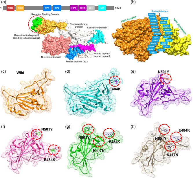
Structural representation of the spike glycoprotein (PDB ID:6M0J) and the receptor‐binding domain of the SARS‐CoV‐2. (a) shows the distribution of different domains differentiated with different colors. The RBD domain is shown as yellow specifically. (B) shows the binding interface of the ACE2 and spike RBD. (c) wild type, (d) E484K, (e) N501Y, and (f) E484K‐N501Y, (g) K417N‐E484K‐N501Y, and (h) shows the K417T‐E484K‐N501Y structure of the spike RBD