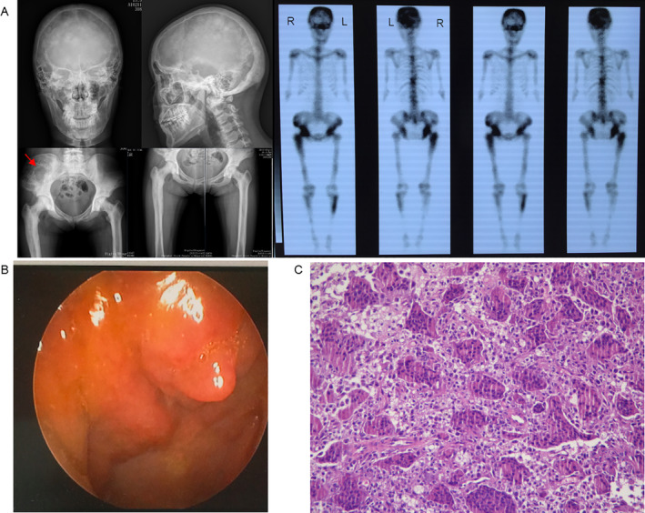
Fig 3.

Clinical features of a patient with sporadic Paget's disease of bone/giant cell tumor of bone (PDB/GCT). (A) X‐ray examination and whole‐body bone scintigraphy of the subject shows radionuclide uptake in the skull, the clavicles, the spine, the pelvis, the humeri, the femurs, and the tibias. X‐ray findings of skull and vertebral body are similar to II‐2 of family 2. The affected long bone showed osteosclerosis and narrow marrow cavity, and large and disordered trabecular bone without osteolysis. The GCT was found at the right ilium. (B) Image of the nasal endoscope, showing tumors on the nasal cavity. (C) Pathological image of tumors resected from the nasal cavity (H&E staining 200×), showing numerous multinucleated giant cells.
