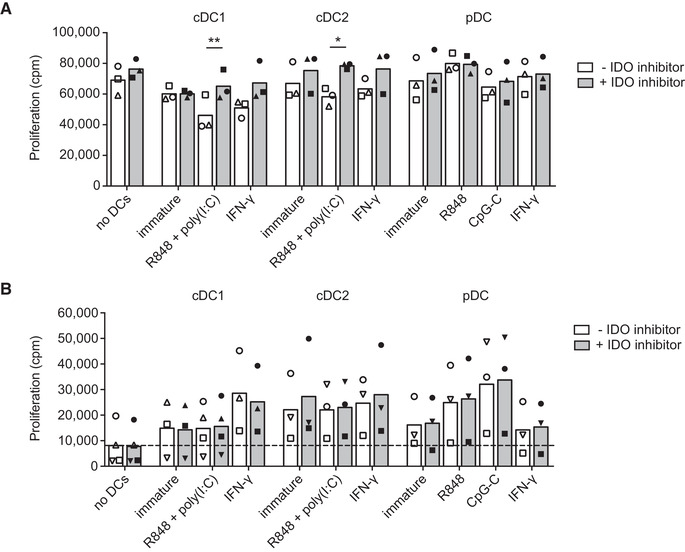
Figure 4.

IDO expressed by cDCs inhibits T cell proliferation. Blood DCs were stimulated with indicated stimuli and/or epacadostat in medium containing 10 μM tryptophan. (A) PBLs were stimulated with anti‐CD3/CD28‐coated beads in supernatants of 48‐hour DC cultures and proliferation was measured after three days by tritiated thymidine incorporation. Mean proliferation in counts per minute (cpm) of three different donors from 3 independent experiments with technical triplicates (cDC2, pDC) or duplicates (cDC1) is shown. Symbols correspond to measurements belonging to the same donors. Significance was determined by two‐tailed paired t‐test comparing absence vs presence of IDO inhibitor (* P < 0.05; ** P < 0.01). (B) Allogeneic PBLs were cultured with overnight‐stimulated DCs and proliferation was measured after another three days by tritiated thymidine incorporation. Mean proliferation of at least three different donors from four independent experiments with technical triplicates (cDC2, pDC) or duplicates (cDC1) are shown. Symbols correspond to measurements belonging to the same donors. Significance was determined by two‐tailed paired t‐test, comparing absence versus presence of IDO inhibitor.
