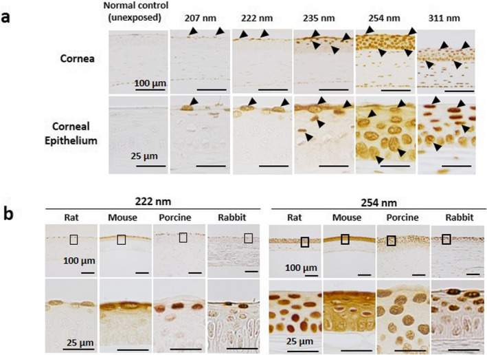
Figure 7.

(a) CPD localization in the cornea immediately after UV irradiation. Arrows indicate PCD‐positive cells. Bar = 100 µm in the upper panel and 25 µm in the lower panel. (b) CPD localization in corneas of the various animals immediately after 222 nm or 254 nm in the UV‐C band. The part surrounded by a black line is indicated below with high magnification. Bar = 100 µm in the upper panel and 25 µm in the high‐magnification images.
