
FIGURE 1.
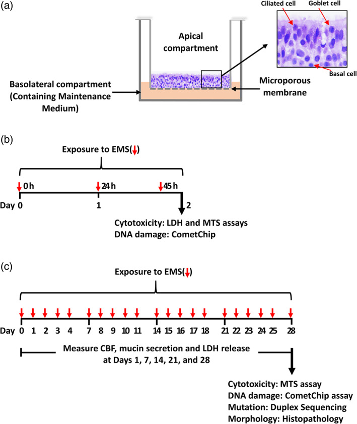
Schematic diagrams of the human ALI airway culture, and experimental design. (a) Shows an image of an H&E stained transverse section from a human ALI tissue model that was used in this study. A microporous membrane that provides the permeable support for cell growth, divides the culture plate well into apical (upper) and basolateral (lower) compartments. The human ALI airway culture consists of ciliated cells, goblet cells, and basal cells. The apical side of the cultures is exposed to the air and Maintenance Medium is added to the basolateral compartment. The cultures were treated with various concentrations of EMS for periods of (b) 3‐days or (c) 28‐days. LDH release, cilia beating frequency and mucin secretion were monitored at Day 1, 7, 14, 21 and 28 of the 28‐day treatment period in (c). The CometChip and cell viability assays were conducted following the 3‐day treatment in (b) and the 28‐day treatment in (c). Cultures were collected for Duplex Sequencing or for morphological investigation at Day 28 in (c). Shorter red arrows: dosed medium added at 0, 24 and 45 h in (b) and Monday through Friday for 28 days in (c); longer arrows: assays performed 3 h after the last treatment (b and c). LDH, lactate dehydrogenase; CBF, cilia beating frequency