
FIGURE 4.
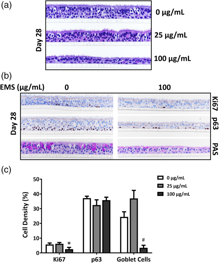
Histological and immunohistochemical (IHC) observations in EMS‐treated ALI airway cultures. (a) Shows representative images of H&E‐stained ALI airway cultures after a 28‐day treatment with EMS. Magnification is 40×. (b) Shows representative images of ALI airway cultures treated with EMS for 28 days and stained for histological and IHC observations. Magnification is 40×. Cell proliferation was evaluated by IHC with anti‐Ki67 antibody; basal cells were stained with anti‐p63 antibody and goblet cells were stained using periodic acid Schiff (PAS). (c) Shows percentages of PAS‐positive goblet cells, anti‐p63‐positive basal cells and anti‐Ki67‐positive proliferating cells. Data (n = 3) presented as means ± SD. *,# p < .05 was considered statistically significant compared to the concurrent vehicle controls; different symbols refer to comparisons made to different controls. A table with primary data has been included as Supporting Information