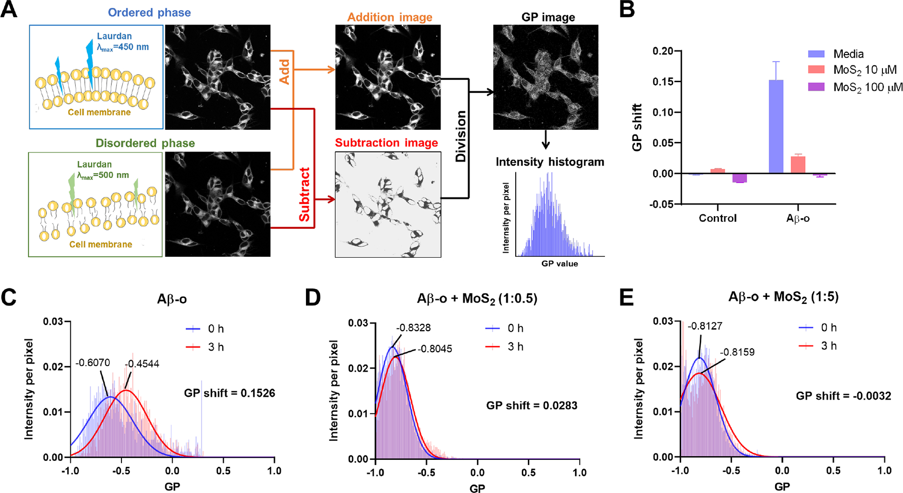
Figure 3. Effect of Aβ oligomers on the fluidity of SH-SY5Y cell membranes in the presence and absence of ultrasmall MoS2 QDs.

(A) A flowchart illustrates calculation of generalized polarization (GP) values from raw ratiometric confocal images in the ordered and disordered phases. The lipid order of cell membranes was indicated by the lipophilic Laurdan dye, which could partition into cell membranes. When the cell membrane was in the liquid ordered phase, Laurdan dye emitted fluorescence at 450 nm under the excitation of 405 nm, and redshifted to 500 nm when the cell membrane was in the liquid disordered phase. GP images and the pixels of cell membranes were derived with ImageJ software. Intensity shifts between the ordered and disordered channels were quantified as GP values. (B) GP shifts were recorded after a 3 h-treatment by Aβ-o (20 μM) in the presence and absence of ultrasmall MoS2 QDs (10 or 100 μM). Ultrasmall MoS2 QDs themselves did not affect cell membrane fluidity in 3 h. (C) Compared to Aβ monomers (Aβ-m) and amyloid fibrils (Aβ-f, Figure S8), Aβ-o are predicted to cause lipid order, and a corresponding positive GP shift. (D, E) MoS2 could significantly prevent damage to cell membrane fluidity caused by Aβ-o and restore the GP values to control cell level at the higher concentration of 100 μM MoS2 QDs.
