Figure 1.

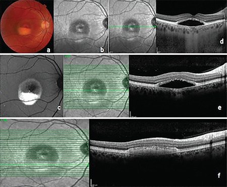
Right eye. a) Color fundus photograph, pseudohypopyon stage; b) Infrared photograph; c) Fundus autofluorescence image; d) Enhanced depth imaging optical coherence tomography (EDI-OCT); e, f) Spectral domain OCT

Right eye. a) Color fundus photograph, pseudohypopyon stage; b) Infrared photograph; c) Fundus autofluorescence image; d) Enhanced depth imaging optical coherence tomography (EDI-OCT); e, f) Spectral domain OCT