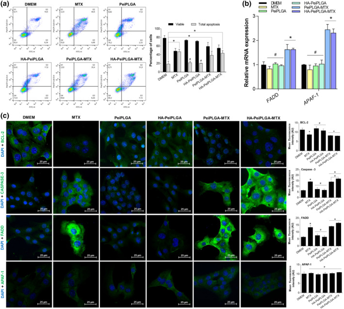
FIGURE 3.

Effect of naked or HA‐coated PeiPLGA‐MTX NPs on apoptosis. The cell death profile was analysed by flow cytometry (a), whose graphical representation expresses the percentage of viable and apoptosis cells. The gene expression profile (b), as well as immunofluorescence immunostaining (c) of key markers in apoptosis were also evaluated. All data are presented as mean ± SD of five independent assays with at least three replicates. *, P < .05, significantly different as indicated; #not significantly different; one‐way ANOVA with post hoc Bonferroni correction or Kruskal–Wallis with post hoc Dunn correction
