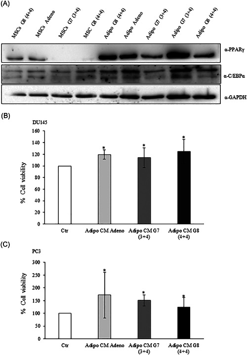
Figure 1.

Adipocyte‐conditioned media effect on prostate cancer cell viability. (A) Mesenchymal stem cells from PPAT were isolated and differentiated as described in Section 2. The lysates were analyzed by immunoblotting with PPARγ and C/EBPα antibodies and autoradiography. GAPDH antibody was used for normalization. (B) DU145 and (C) PC3 (2 × 10⁴ cells) cells were plated in 96 well plates and serum starved for 16 h cells. Then, the cells were incubated with 0,25% BSA or PPAT Adipocyte‐CM from Adeno, G7(3 + 4) and G8(4 + 4) adipocytes for 48 h. Cell viability was assessed by the MTT assay. The results were reported as percentage of viable cells compared to control, considered as maximum viability (100%). Data represent the mean ± SD of triplicate samples of three independent experiments. The bars represent the mean ± SD of at least three independent experiments. *p value < .05. BSA, bovine serum albumin; C/EBPα, CCAAT/enhancer‐binding protein alpha; MTT, 3‐(4,5‐dimethylthiazol‐2‐yl)‐2,5‐diphenyltetrazolium bromide; PPARγ, peroxisome proliferator‐activated receptor gamma; PPAT, periprostatic adipose tissue; SD, standard deviation
