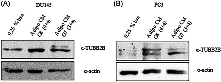
Figure 3.

TUBB2B expression in prostate cancer cell. (A) DU145 and (B) PC3 were serum starved for 16 h cells and then incubated with 0,25% BSA or PPAT Adipocyte‐CM from G7(3 + 4) and G8(4 + 4) adipocytes for 48 h. The lysates were analyzed by immunoblotting with TUBB2B antibody and autoradiography. Actin antibody was used for normalization. The autoradiograph shown is representative of three different experiments. BSA, bovine serum albumin; PPAT, periprostatic adipose tissue; TUBB2B, β‐tubulin isoform 2B
