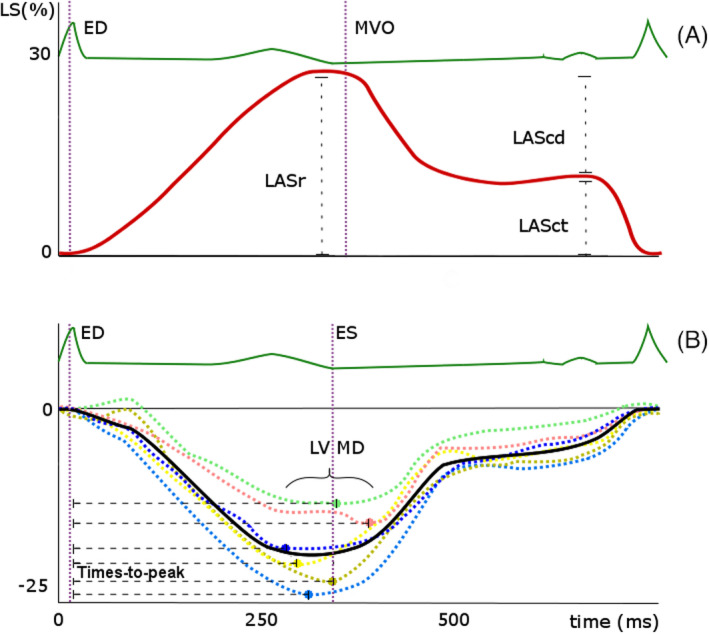
FIGURE 1.

(A) Typical curve of left atrial (LA) longitudinal strain (LS), which has a positive value. End‐diastole (ED) is set at the QRS‐peak of the ECG, peak reservoir strain (LASr) typically precedes mitral valve opening (MVO). Passive (LAScd; conduit strain) and active (LASct; contractile strain) left atrial emptying are seen during ventricular diastole (B) Conceptualization of left ventricular mechanical dispersion (LV MD), which is the standard deviation of the 18 segmental time intervals from the QRS‐peak on the ECG to peak negative strain. Dashed curves are segmental longitudinal strain (LS) curves, the solid curve is the global longitudinal strain curve. ED =end‐diastole, ES =end‐systole (aortic valve closure, for which the surrogate of minimal volume is used)
