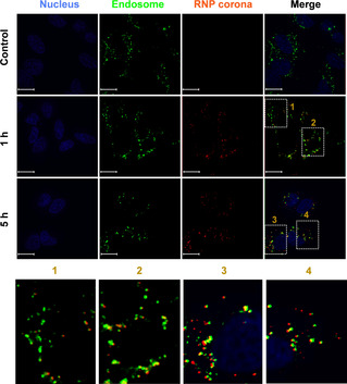
Figure 3.

Endocytosis of the binding‐mediated RNP corona. See Figure S12 for experimental details. The endosomes/lysosomes were stained with LysoTracker Yellow HCK‐123 (50 nM, green) and the nuclei were stained with DAPI (4,6‐diamidino‐2‐phenylindole, blue). The RNP corona was red, due to the Cy5 labeling. Scale bar=15 μm.
