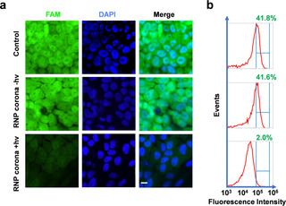
Figure 4.

Editing of the EGFP gene in Hela‐GFP cells using the RNP corona system. a) Confocal microscopy images of Hela‐GFP cells. Cells were treated with RNP corona (ratio of 10:1, 10 nM) for 8 h, and irradiated at 3.0 mW cm−2 or kept in the dark for 15 min. The loss of fluorescence was measured five days after treatment. Scale bar=10 μm. b) Flow cytometry histograms of the Hela‐GFP cells corresponding to treatment in (a).
