
Figure 3.
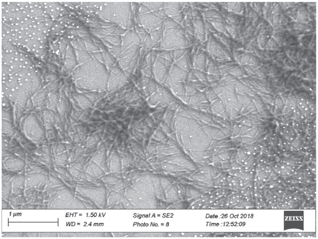
Scanning electron microscopic image of an insulin fibril network measured with a ZEISS Scanning Electron Micro-scope (Sigma 300 VP, Oberkochen, Germany). The white dots are gold nanoparticles from the sputtering process.
Official websites use .gov
A
.gov website belongs to an official
government organization in the United States.
Secure .gov websites use HTTPS
A lock (
) or https:// means you've safely
connected to the .gov website. Share sensitive
information only on official, secure websites.
Scanning electron microscopic image of an insulin fibril network measured with a ZEISS Scanning Electron Micro-scope (Sigma 300 VP, Oberkochen, Germany). The white dots are gold nanoparticles from the sputtering process.