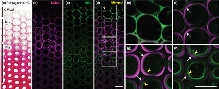
Fig. 4.

Simultaneous visualisation of laccase‐dependent DMAC‐PA and NBD‐CA incorporation into differentiating compression wood tracheids of Chamaecyparis obtusa seedlings. (a) A control section stained with phloroglucinol‐HCl. (b–h) Sections were labelled sequentially with DMAC‐tagged p‐coumaryl alcohol (DMAC‐PA) and NBD‐tagged coniferyl alcohol (NBD‐CA) probes in the presence of catalase, and then visualised as DMAC (b) and NBD (c) fluorescence and merged (d–h). Images in (e–h) are magnified views of the boxed regions in (d). White arrows and yellow arrowheads indicate the S2L and iS2 layers, respectively. Regions where the incorporation of fluorescence‐tagged monolignols was detected in different cell wall compartments are indicated in (a). Bars, 20 μm.
