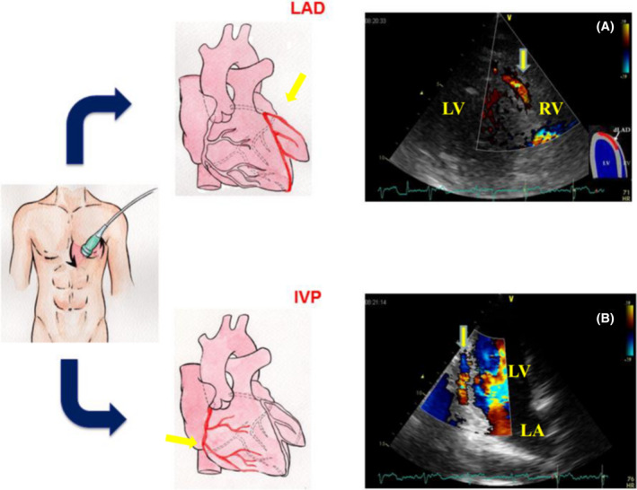
FIGURE 1.

Color Doppler images of the distal left anterior descending coronary artery (LAD) and interventricular posterior coronary artery (IVP). The modified apical 4‐chamber (A) and 2‐chamber (B) positions, with cranial angulation of the transducer and a Nyquist limit of 20‐67 cm/s, allow optimal identification of the diastolic coronary flow. LV = left ventricle; RV = right ventricle; LA = left atrium
