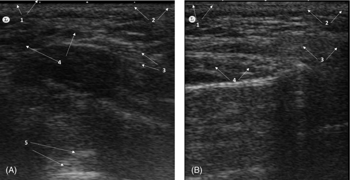
FIGURE 3.

Heterogeneous pattern of a patient injected with hyaluronic acid filler. (A) Immediately after treatment. Poorly defined globular ultrasound pattern, with anechoic images indicative of liquid content. 1: Epidermis; 2: dermis; 3: Subcutaneous cellular tissue; 4: anechoic images; 5: Posterior echogenic reinforcement. (B) 1 month after treatment. Typical heterogeneous pattern, without residual anechoic areas, which was indicative of a total integration of Hyaluronic acid filler. 1. Epidermis; 2: Dermis; 3: Subcutaneous cellular tissue; 4: Heterogeneous pattern
