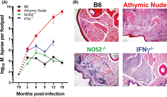
FIGURE 1.

Growth of M. leprae and granulomatous response in infected footpads. A, B6, athymic nude, NOS2−/−, and IFN‐γ−/− mice were infected in the hind footpads with 6 × 10³ M. leprae, and growth was monitored for 18 mo. B, Histological examination (hematoxylin and eosin stains) of M. leprae–infected footpads at 9 mo postinfection. B6 develop a small granuloma comprised of lymphocytes, epithelioid cells, and macrophages. Athymic nude mouse footpads become engorged with M. leprae filled macrophages. In NOS2−/− mouse footpads, the large granulomatous response infiltrated the perineurium and muscle bundles, whereas in IFN‐γ−/− footpads, the large unorganized infiltration was composed of epithelioid macrophages with randomly interspersed lymphocytes
