Fig 3.

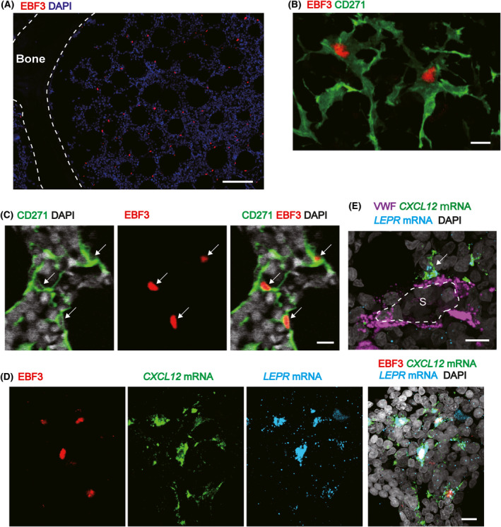
CXCL12hi cells are identified by EBF3 staining in human adult marrow sections. (A) Immunohistochemical analysis for EBF3 in human adult bone marrow. EBF3+ cells were scattered throughout the bone marrow cavity. (B) Immunohistochemical analysis for EBF3 and CD271 in human adult bone marrow. Membranes of EBF3+ cells were positive for CD271, and EBF3+CD271+ cells displayed long processes that formed a reticular network. (C) Immunohistochemical analysis for EBF3 and CD271 in human adult bone marrow. Almost all DAPI+ nuclei of CD271+ cells (white arrows) were positive for EBF3 in the marrow cavity. (D) Combined in situ hybridization of the CXCL12 and LEPR mRNAs and immunohistochemistry for EBF3 in human adult bone marrow. (E) Combined in situ hybridization of the CXCL12 and LEPR mRNAs and immunohistochemistry for von Willebrand factor (VWF) in human adult bone marrow. Cells expressing CXCL12 (white arrow) in contact with endothelial cells (ECs) of vascular sinuses (S) were shown. Scale bar: (A) 100 µm; (B) 10 µm; (C) 10 µm; (D) 10 µm; (E) 10 µm.
