
FIGURE 3.
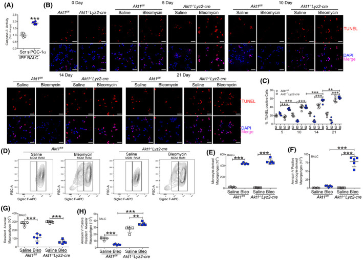
Monocyte‐derived macrophages are resistant to apoptosis. A, Caspase‐3 activity (n = 4‐6) in IPF BALC with silencing of PGC‐1α. BAL cells were isolated at indicated days after saline (n = 5/time point) or bleomycin exposure (n = 5/time point) from Akt1fl/fl or Akt1−/−Lyz2‐cre mice. B, TUNEL staining (n = 5) and C, quantification (n = 5). Scale bar represents 40 μm. BAL cells were isolated 21 days after exposure. D, Representative flow cytometry plots of monocyte‐derived macrophages (MDM, CD45+CD11b+/−Ly6G−CD64+Ly6c−Siglec Flow) and resident alveolar macrophages (RAM, CD45+CD11b+/−Ly6G−CD64+Ly6c−Siglec Fhi) from saline or bleomycin‐exposed Akt1fl/fl and Akt1−/−Lyz2‐cre mice. Total cell number of (E) MDM (n = 5), F, annexin V positive MDM (n = 5), (G) RAM (n = 5), and H, annexin V positive RAM from Akt1fl/fl and Akt1−/−Lyz2‐cre BAL (n = 5). **P < .001; ***P < .0001. Values shown as mean ± S.E.M. Two‐tailed t‐test statistical analysis was utilized for A. One‐way ANOVA followed by Tukey’s multiple comparison test was utilized for C, E‐H