
FIGURE 4.
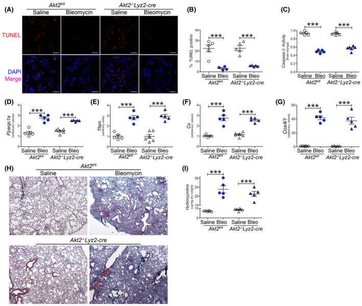
Pulmonary fibrosis does not require Akt2. Akt2fl/fl or Akt2−/−Lyz2‐cre mice were exposed to saline or bleomycin (Bleo). BAL cells were isolated 21 days later. A, TUNEL staining (n = 5), B, TUNEL quantification (n = 4‐5), and C, caspase‐3 activity (n = 5). Scale bar represent 50 μm. mRNA analysis of D, Ppargc1a (n = 5‐6), E, Tfam (n = 5‐6), F, Cs (n = 5‐6), and G, Cox4i1 expression (n = 5‐6) in BAL cells. H, Histology of lung sections with Masson’s trichrome staining (n = 5) and I, hydroxyproline analysis of homogenized lung (n = 5‐6). ***P < .0001. Values shown as mean ± S.E.M. One‐way ANOVA followed by Tukey’s multiple comparison test was utilized