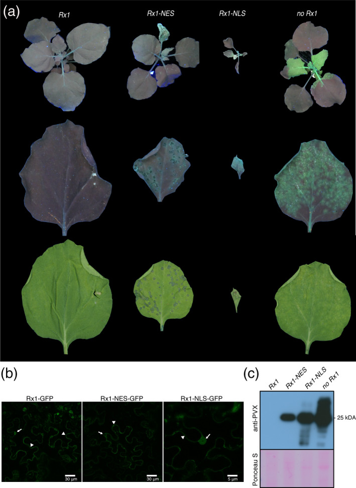
Figure 1.

Rx1 localization variants Rx1‐NLS and Rx1‐NES failed to block PVX‐GFP infection. (a) PVX rub‐inoculated 5‐week‐old Nicotiana benthamiana plants at 8 days post‐inoculation (8 dpi) under UV light, and a PVX‐GFP rub‐inoculated leaf under UV and white light at 6 dpi, presented in top, middle and bottom rows, respectively. (b) Whereas Rx1 shows a nuclear–cytosolic distribution, the variants are either nuclear excluded (Rx1‐NES) or nuclear enriched (Rx1‐NLS). Subcellular localization of GFP‐tagged Rx1 and Rx1 variants visualized by confocal microscopy. The 35SLS::Rx1‐GFP, 35SLS::Rx1‐NLS‐GFP and 35SLS::Rx1‐NES‐GFP constructs where transiently expressed in N. benthamiana. Arrows and arrowheads indicate the nucleus and the cytoplasm, respectively. (c) Immunodetection of PVX‐GFP in systemic leaves of rub‐inoculated N. benthamiana transgenic lines expressing Rx1, Rx1‐NES and Rx1‐NLS, 8 days after rub‐inoculation. The immunoblotting was performed using polyclonal anti‐PVX antibody followed by incubation with horseradish peroxidase (HRP)‐conjugated goat anti‐rabbit immunoglobulin G (IgG) secondary antibody. Ponceau S staining shows the equal protein loading of the samples.
