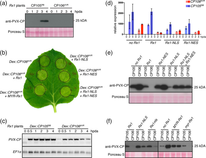
Figure 2.

Rx1, Rx1‐NES, Rx1‐NLS or the combination of Rx1‐NES plus Rx1‐NLS trigger a hypersensitive response (HR) upon CP106AVR (Avirulent) expression, but only wild‐type (WT) Rx1 is able to prevent CP106AVR protein accumulation. (a) Detection of CP105RB (Resistance Breaking) and CP106AVR proteins in Rx1 Nicotiana benthamiana plants after dexamethasone (Dex) induction, by Western blot. Ponceau S staining shows equal protein loading. (b) HR after co‐expression of Rx1 localization variants and Dex::CP106AVR 1 day post Dex induction (1 dpda). Circles depict the infiltrated zones containing the Agrobacterium tumefaciens strains carrying the indicated constructs. (c) Detection of CP105RB and CP106AVR transcripts in Rx1 N. benthamiana plants after Dex induction, by semi‐quantitative RT‐PCR. The EF1α control serves as a control for equal quantities of mRNAs used in the semi‐quantitative RT‐PCR. (d) CP105RB and CP106AVR transcript levels were quantified by RT‐qPCR at 2 and 4 hpda, relative to 0 hpda, in plants co‐expressing different Rx1 constructs. For each data point, the cycle threshold (C t) values of three replicates were normalized to the C t values obtained for the reference genes EF1α and PP2A using the 2−ΔΔ C t method. (e) Detection of coat protein (CP) by Western blot in N. benthamiana plants co‐expressing different Rx1 localization variants (NLS, nuclear localization signal; NES, nuclear export signal) with either Dex::CP105RB or Dex::CP106AVR, at 4 hpda. (f) Detection of CP by Western blot in N. benthamiana co‐expressing different Rx1 localization variants. CP106AVR does not accumulate in the presence of Rx1 or in the presence of Rx1 variants carrying a mutant NLS (Rx1‐nls) or myristoylation motif (myr‐Rx1). CP106AVR accumulates when Rx1 is sequestered in the nucleus (Rx1‐NLS) or tethered at the plasma membrane (MYR‐Rx1). For PVX‐CP CP105RB or CP106AVR by Western blot. For immunoblotting, proteins were extracted from N. benthamiana leaves at 4 hpda, unless otherwise specified. The immunoblotting was performed using polyclonal anti‐PVX antibody followed by incubation with horseradish peroxidase (HRP)‐conjugated goat anti‐rabbit immunoglobulin G (IgG) secondary antibody. The photosensitive film was exposed to the membrane 2 min before developing.
