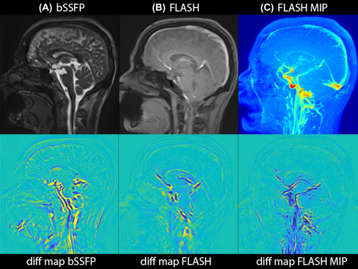
FIGURE 7.

3D aMRI applied to FLASH, demonstrating the applicability of the 3D aMRI algorithm to other MR contrasts. Maximum difference maps calculated from 3D aMRI are shown for volumetric (bSSFP) data (A) and 2D FLASH cine data (B). C, An MIP of the amplified FLASH data is shown together with its corresponding difference map, allowing one to visualize the pulsation of the major blood vessels
