
Fig 8.
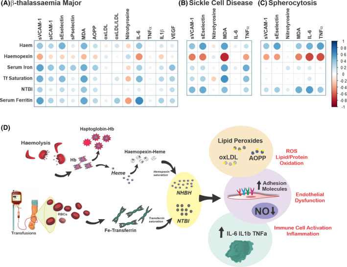
Working model: haem‐ and iron‐mediated toxicity in haemolytic diseases. (A) Correlation plots between haem/iron‐related parameters [serum haem, iron, transferrin (Tf) saturation, non‐Tf‐bound iron (NTBI), haemopexin (Hx)] and damage‐related parameters [malondialdehyde (MDA), soluble vascular cell and intercellular adhesion molecules 1 (sVCAM1, sICAM‐1), soluble endothelial and platelet selectins (sP‐selectin, sE‐selectin), oxidation of low‐density lipoprotein (oxLDL), nitrotyrosine, advanced oxidised protein product (AOPP), interleukin 1β (IL‐1β), tumour necrosis factor α (TNFα), IL‐6, vascular endothelial growth factor (VEGF)] in patients with β‐thalassaemia (β‐thal) major. Corresponding Spearman correlation coefficients and P values are reported in Table SIII. (B, C) Correlation plots between iron‐related parameters (haem, iron, Tf saturation, NTBI, Hx) and damage‐related parameters [sVCAM1, sE‐selectin, nitrotyrosine, MDA, IL‐6, TNFα, VEGF, monocyte chemoattractant protein‐1 (MCP‐1)] in patients with sickle cell disease (SCD; SCD 1 and 2)and spherocytosis (SPH). Corresponding Spearman correlation coefficients and P values are reported in Tables SIX and SX. The size of the dot suggests the strength of the correlation; the colour of the dot indicate the direction of the correlation (blue dot: positive; red dot: negative). (D) In haemolytic diseases, haemolysis leads to the release of elevated haemoglobin (Hb) and haem levels in the circulation. Saturation of the Hb and haem scavengers haptoglobin and haemopexin triggers the formation of ‘free’ non‐haptoglobin‐bound Hb (NHBHb), non‐haemopexin‐bound haem (NHBH). In parallel, transfusions increase systemic iron levels and transferrin saturation. This generates ‘free’ NTBI. NHBHb, NHBH and NTBI exert a synergistic detrimental action on the vasculature and immune cells. While the pro‐oxidant potential of NHBH and NTBI is reflected by elevated lipid and proteins oxidation (lipid peroxides, oxLDL, AOPP), their vasculo‐toxic and pro‐inflammatory action is reflected by elevated soluble adhesion molecules (e.g. E‐selectin, P‐selectin, ICAM‐1, VCAM‐1), VEGF and inflammatory cytokines (e.g. IL‐6, TNFα, IL‐1β) and reduced nitric oxide (NO) levels. These alterations likely contribute to NHBH‐ and NTBI‐triggered vasculopathy and chronic sterile inflammation, typical hallmarks of several haemolytic diseases. [Colour figure can be viewed at wileyonlinelibrary.com]