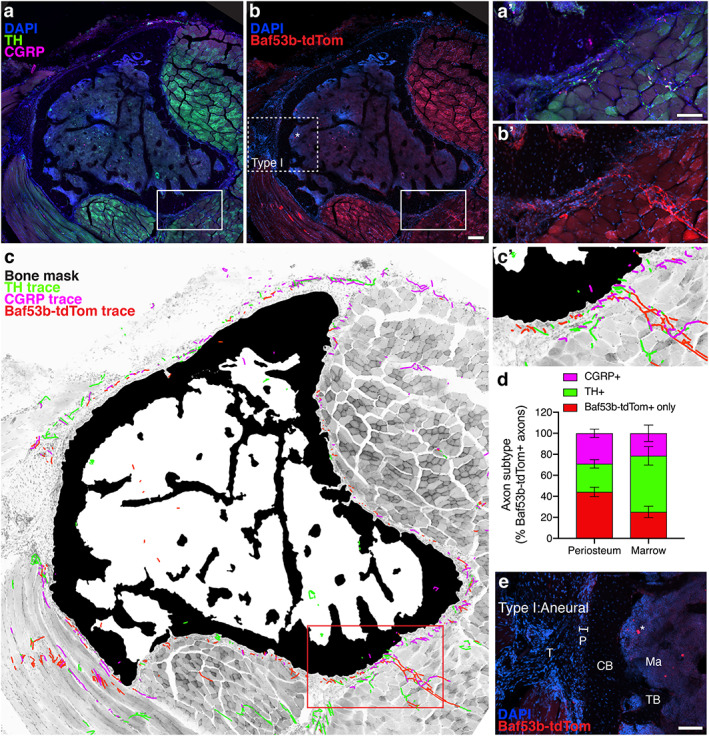
Fig 3.

Baf53b‐tdTomato is a robust, selective reporter system for skeletal innervation. (A, B) Representative confocal micrograph of a proximal tibial cross section with immunolabeled CGRP+ sensory axons (magenta) and TH+ sympathetic axons (green) with DAPI+ nuclei (blue, A), and tdTomato expression driven by Baf53b‐Cre, as well as DAPI+ nuclei (blue, B). Asterisks indicate non‐neural, autofluorescence. Scale bar = 200 μm. (C) Bone mask (black), CGRP+ axon traces (magenta), TH+ axon traces (green), and Baf53b‐tdTomato + axon traces overlaid on a grayscale max projection to visualize tibial innervation. CGRP+ and TH+ axons were also positive for Baf53b‐tdTomato. (A′–C′) High magnification of the periosteal innervation of the solid boxed region indicated in A–C. Scale bar = 100 μm. (D) Quantification of compartmentalized axonal subtypes pooled from L1 to L4 represented as a percentage of total Baf53b‐tdTomato + axons. Bars represent mean with SEM; n = 3 individual animals quantified at each level. (E) Aneural region of the periosteum (P), where tendon (T) connects to cortical bone (CB), is devoid of Baf53b‐tdTomato+ axons. TB = trabecular bone; ma = marrow. Scale bar = 100 μm.
