Fig 5.

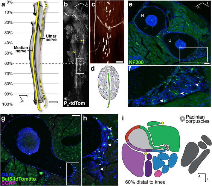
Specialized mechanoreceptor endings are associated with the skeleton of the forearm and lower leg. (A) Schematic of major nerve branches in the forearm overlaid on a 3D‐rendered μCT scan of the radius–ulna. Scale bar = 1 mm. (B) Light‐sheet image of a cleared forearm from an adult Schwann cell reporter mouse (P0‐tdTomato) revealing the ulnar nerve (filled arrowhead) and median nerve (empty arrowhead) and the distribution of Pacinian corpuscles on the ulnar surface. (C) High‐magnification, intensity‐pseudocolored image of the boxed region in B. Scale bar = 200 μm. (D) Drawing of a Pacinian corpuscle demonstrating the lamellar structure derived from Schwann cells surrounding a single unmyelinated ending of a large, myelinated sensory axon. (E) Representative confocal micrograph through a 100‐μm‐thick transverse cross section through the radius (R) and ulna (U) and surrounding musculature visualized by DAPI (blue) and NF200 (green) immunolabeling at 60% of the ulnar length. Scale bar = 200 μm. (F) High magnification of the corpuscles (arrowheads) on the lateral aspect of the ulna of the boxed region indicated in E. (G) Representative confocal projection of a 50‐μm‐thick transverse cross section of a tibia from a Baf53b‐tdTomato (green) reporter mouse with immunolabeling CGRP+ axons (magenta) along with DAPI staining (blue) at 60% of the tibial length distal to the knee. Scale bar = 200 μm. (H) High magnification of the Pacinian corpuscles on the posterior aspect of the tibia of the boxed region indicated in G. (I) 2D map corresponding to the confocal image in G indicating the location of Pacinian corpuscles found in all strains and sexes at this level of the tibia. Additional information can be found in the supplemental tibial atlases (Supplemental Files C, D).
