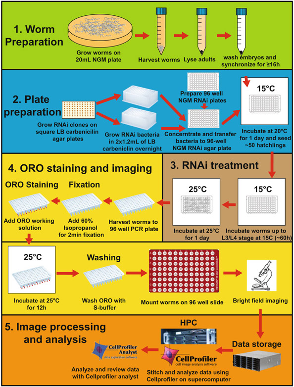
Fig. 1.
RNAi screening workflow. Worm preparation is shown in green (see Subheading 3.3); bacterial RNAi preparation is shown in blue (see Subheading 3.2); worm RNAi treatment is shown in brown (see Subheading 3.4); ORO staining and imaging procedures are shown in yellow (see Subheadings 3.5 and 3.6); and image analysis and data transfer and processing are shown in orange (see Subheading 4)

