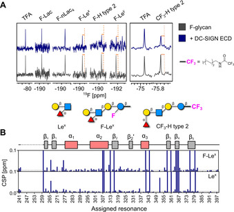
Figure 2.

Mammalian lectin (DC‐SIGN) binding to F‐glycans and study on the reporter position. A) CPMG NMR screening of F‐glycans alone (gray) and in presence of DC‐SIGN ECD (blue). DC‐SIGN ECD binds to F‐Lex, F‐H type 2, and F‐Ley as shown by a decrease in peak intensity in presence of protein (orange lines, left panel). CPMG NMR spectra of CF3‐H type 2 alone (gray) and in presence of DC‐SIGN ECD (blue; right panel). B) Cartoon of assigned domains of DC‐SIGN CRD (unassigned resonances in dashed line) and CSP plot of assigned resonances in presence of F‐Lex and Lex showing that F‐Lex‐perturbed resonances similarly to unlabeled Lex.
