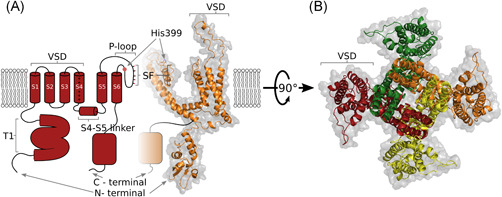
Figure 1.

Structural representations of the KV1.3 channel. (A) Hybrid side view as two‐dimensional (left, red) and three‐dimensional homology (right, orange) representations of two opposing domains in (B). (B) Top (extracellular) view of all four domains, with each colour coded, which shows the domain‐swapping architecture of the KV1.3 channel. The homology model was built using Modeller 9.21, Kv1.2 (PDB 3LUT) as a template, Kv1.3 sequance was retrieved from Uniprot (Accession No.: P22001), and the figure was prepared using PyMOL70, 71, 72, 73 [Color figure can be viewed at wileyonlinelibrary.com]
