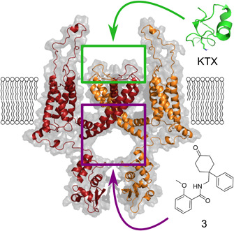
Figure 3.

Representation of the binding sites of KV1.3 inhibitors. Green square, extracellular binding site of KTX toxin (green, PDB 1KTX) as a representative toxin; purple square, central cavity preserved in many K+ ion channels as the binding site for small molecules, such as believed for inhibitor 3. The homology model was build using Modeller 9.21, Kv1.2 (PDB 3LUT) as a template, KV1.3 sequence was retrieved from Uniprot (Accession No.: P22001), and the figure was prepared using Pymol70, 71, 72, 73 [Color figure can be viewed at wileyonlinelibrary.com]
