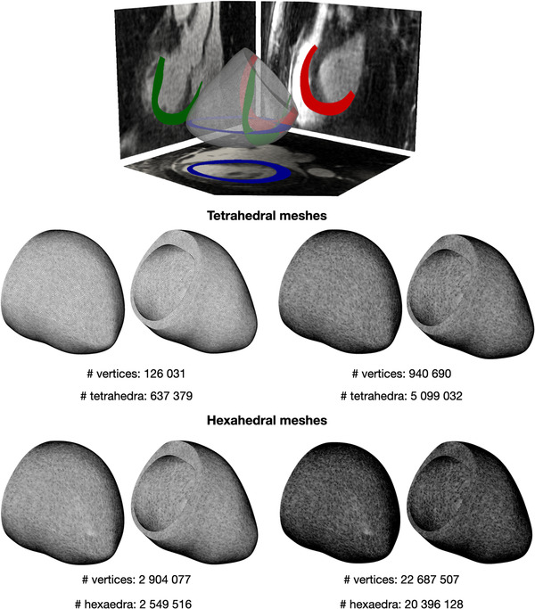
FIGURE 2.

Two tetrahedral (center) and two hexahedral (bottom) meshes with different levels of refinement. Geometry was obtained from segmentation of patient specific MRI images (top image from ref. 40) [Color figure can be viewed at wileyonlinelibrary.com]
