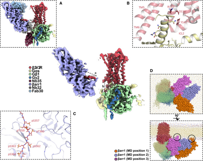
Fig 3.

(center) Structure of the β2V2R–Gs protein–βarr1 megaplex with all stabilizing protein components removed. (A) Same as in center, but with all stabilizing proteins shown. (B) Interaction between the Gs α5 helix of Gs protein and the β2V2R. Critical receptor residues within the DRY motif and ICL2 are labeled. (C) Binding interface between the phosphorylated β2V2R tail (V2T) and βarr1, with phosphorylated V2T residues labeled. (D) Orthogonal views of the final frame of a coarse‐grained molecular dynamics simulation of a megaplex with three coarse‐grained models that each differ in their βarr1 position relative to the β2V2R–Gs protein portion of the megaplex. Circles indicate contacts observed between the βarr1 C‐edge loops with the membrane.
