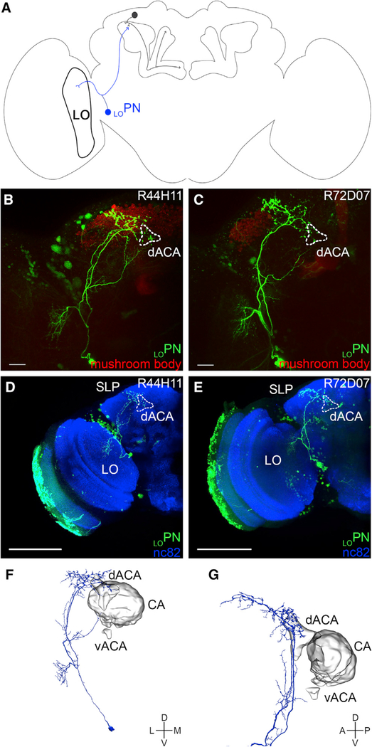
Figure 4. LOPN Connecting the Lobula to the dACA.
(A) A schematic of the Drosophila brain shows an α/βp Kenyon cell input neuron—LOPN (blue)—projecting from the lobula (LO) to the dACA.
(B and C) LOPN (bright green) was identified in the screen using two different transgenic lines (B: R44H11-GAL4 and C: R72D07-GAL4); the neurons photo-labeled in each line show an overall similar morphology.
(D and E) LOPN was photo-labeled using either the R44H11-GAL4 (D) or R72D07-GAL4 (E) transgenic lines; the samples were fixed, immuno-stained (nc82 antibody, blue), and imaged. The photo-labeled neurons show an overall similar morphology: their somata are located near the optic lobe; they extend dendritic terminals in a small region of the LO; and they extend axonal terminals in the dACA and the SLP.
(F and G) A LOPN-like neuron was identified in the hemibrain connectome: (F) this neuron projects from the LO to the dACA and the SLP and (G) its axonal terminals innervate the dACA, but not the CA or the vACA. The images in (F) and (G) were taken directly from the Neuprint platform. The neuropil domains are defined by Neuprint platform.
The following genotypes were used in this figure: (B and D) yw/yw;MB247-DsRedunknown,UAS-C3PA-GFPunknown/UAS-C3PA-GFPattP40;UAS-C3PA-GFPattP2,UAS-C3PA-GFPVK00005,UAS-C3PA-GFPVK00027/R44H11-GAL4attP2 and (C and E) yw/yw;MB247-DsRedunknown,UAS-C3PA-GFPattP40/UAS-C3PA-GFPunknown;UAS-C3PA-GFPattP2,UAS-C3PA-GFPVK00005,UAS-C3PA-GFPVK00027/R72D07-GAL4-GFPattP2. Scale bars are 20 μm (B and C) and 100 μm (D and E).

