Figure 1.
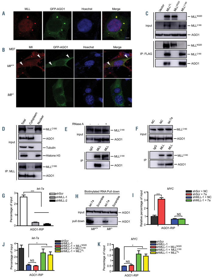
MLL is required for the loading of let-7a onto AGO1. (A) 293T cells were transfected with GFP-AGO1. Immunofluorescence experiments were performed to visualize the localization of GFP-AGO1 and MLL. MLL-CT antibody, which recognizes MLLC180 (aa2829-2883), was used to detect MLL. Scale bar, 5 mm. (B) Mll wild-type (Mll+/+) and Mll knockout (Mll-/-) MEF cells were transfected with GFP-AGO1. Immunofluorescence experiments were performed to visualize the localization of GFP-AGO1 and MLL. Arrowheads show the localization of MLL with the GFP-AGO1. Scale bar, 5 mm. (C) 293T cells were transfected with FLAGtagged full-length MLL (MLLFL), MLLC320, MLLC180 or empty vector. Cell lysates were prepared and subjected to anti-FLAG immunoprecipitation assays. The interaction between MLL and AGO1 was analyzed by western blot assays using indicated antibodies. (D) The cytosolic and nuclear fractions of 293T cells were separated and subjected to immunoprecipitation using anti-MLL antibodies. Co-purified proteins were examined by immunoblots using the indicated antibodies. (E) 293T cell lysates were treated with RNase A followed by anti-MLL immunoprecipitation. Western blots were performed using the indicated antibodies. (F) The interaction between MLL and AGO1 was assessed after let-7a transfection. Anti-MLL immunoprecipitation assays were performed, results were analyzed by immunoblots with indicated antibodies. (G) Extracts of 293T-shScr and 293T-shMLL cells were subjected to RNA immunoprecipitation (RIP) analysis using anti- AGO1 antibody, and pulled down RNA were analyzed by quantitative reverse transcription polymerase chain reaction (qRT-PCR) using specific primers for let-7a. (H) Mll+/+ and Mll-/- MEF cellular lysates were subjected to a biotinylated- let-7a RNA pull-down assay. Then let-7a-immunoprecipitated AGO1 proteins were subjected to western blot analysis. Scrambled miRNA were used as a negative control. (I) 293T-shScr and 293T-shMLL cells were transfected with Agomir-negative control (NC) and Agomir- let-7a mimic (let-7a) followed by anti-AGO1 RIP experiments at 24 h post-transfection. Total RNAs were isolated to analyze the MYC mRNA level by qRT-PCR. (J, K) 293T-shScr and 293T-shMLL cells with the latter being rescued by exogenous shRNA-resistant MLLN320, MLLC180 or MLLFL were performed with anti-AGO1 RIP experiments at 24 h after transfection. Total RNA were isolated to analyze the let-7a (J) and MYC (K) levels by qRT-PCR using specific primers. NS, no significant difference. *P<0.05, **P<0.01, ***P<0.001. Data represent mean and standard error fo mean of three independent experiments.

