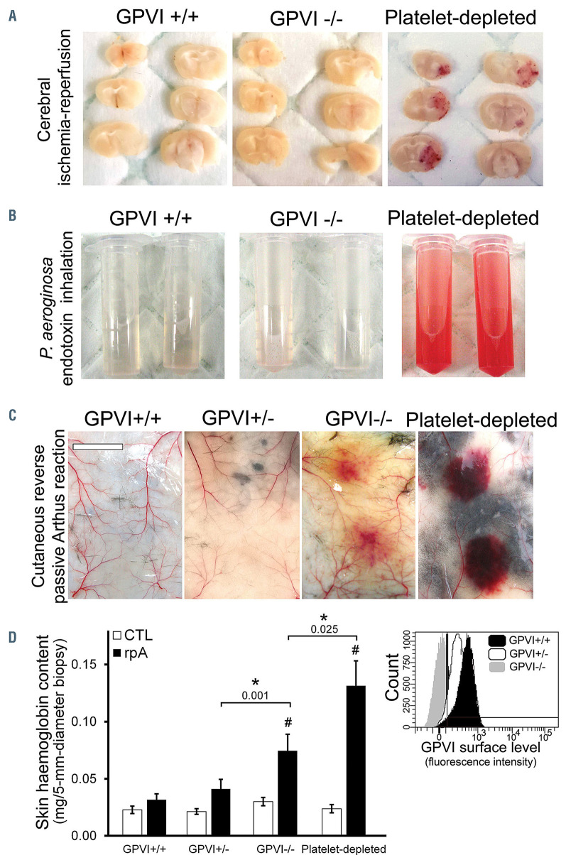
Figure 1.
Contribution of glycoprotein VI to inflammation-associated hemostasis. The contribution of glycoprotein VI (GPVI) to inflammation-associated hemostasis was determined in three different models of acute inflammation. (A) Representative images of brain sections taken 24 hours after GPVI+/+, GPVI-/-, GPVI-/-, and platelet-depleted mice were subjected to 90 minutes transient middle cerebral artery occlusion (tMCAO). Note that tMCA0 caused bleeding only in platelet-depleted mice. The images are representative of n=6 mice per group. (B) Representative images of the bronchoalveolar lavage fluid from Gpvi+/+, Gpvi- /-, and platelet-depleted mice collected 24 hours after lipopolysaccharide inhalation. The images are representative of n=8 mice per group. (C and D) Effect of partial or complete GPVI deficiency on inflammatory bleeding during the cutaneous reverse passive Arthus reaction (rpA). (C) Representative images of the skin of GPVI+/+, GPVI-/-, GPVI-, and platelet-depleted mice after 4 hours of rpA. The images are representative of n=7-10 mice per group. Bar =500 mm. (D) Skin hemoglobin content after 4 hours of rpA. # indicates a significant difference (P<0.05) from the rpA GPVI+/+ group, n=14-20 skin biopsies per group. Inset: Representative histogram of flow cytometry analysis of GPVI surface levels in GPVI+/+, GPVI+/-, and GPVI-/- mice, as assessed using the JAQ1 antibody to representative mouse GPVI.

