Figure 5.
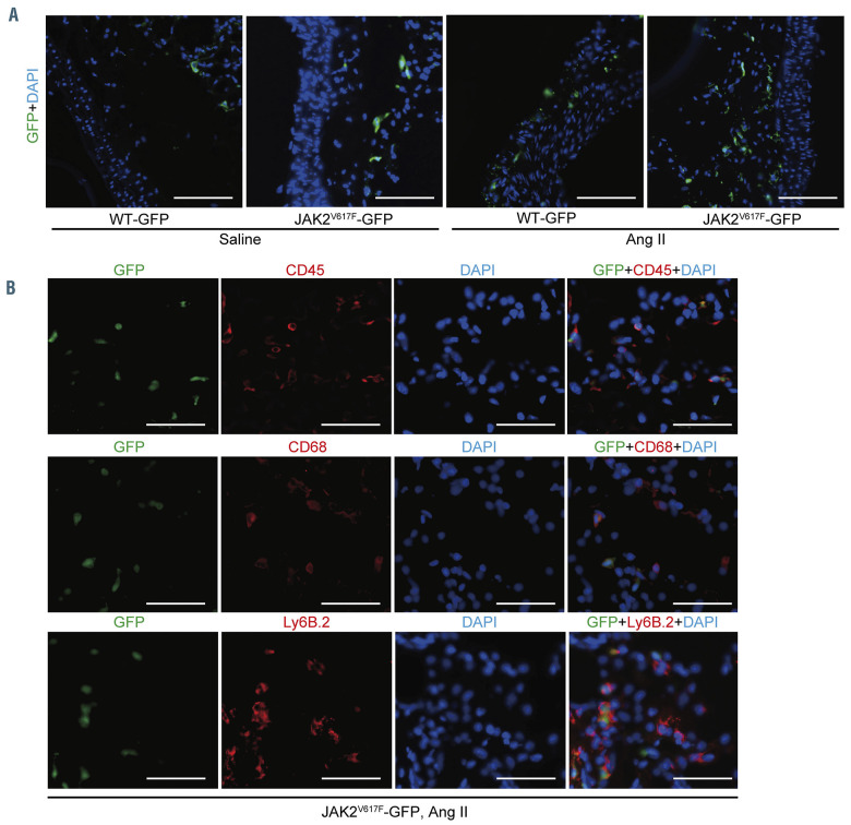
Characterization of bone marrow-derived JAK2 V617F hematopoietic cells in the abdominal aorta by use of GFP-transgene. (A) The lethally irradiated ApoE−/− mice were transplanted with bone marrow (BM) cells from the WT/CAG-EGFP (WT-GFP) mice or JAK2V617F/CAG-EGFP (JAK2V617F-GFP) double transgenic mice. The ApoE−/− recipient mice were subjected to saline or angiotensin II (Ang II) infusion for 4 weeks. The abdominal aortas were stained with an anti-GFP (green) antibody and DAPI (blue). Scale bars, 100 m. (B) Representative immunofluorescence images of the aorta sections stained with anti-CD45 (red), anti-CD68 (red), or anti-Ly6B.2 (red) and anti-GFP (green) antibodies and DAPI (blue) in ApoE−/− mice transplanted with JAK2V617F-GFP BM cells. Scale bars, 50 m. Ang II: angiotensin II; GFP: green fluorescent protein; DAPI: 4′,6-diamidino-2-phenylindole; WT: wild-type.

