Figure 1.
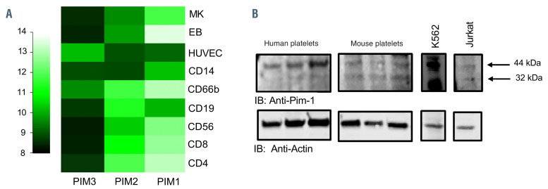
Expression of Pim kinase in human and mouse platelets. (A) HaemAtlas analysis of Pim kinase mRNA expression levels. Pim kinase mRNA levels were quantified in human megakaryocytes and a range of blood cells by analysis of gene array data. Megakaryocytes (MK), human erythroblasts (EB), human umbilical vein endothelial cells (HUVEC), monocytes (CD14), granulocytes (CD66), mature B cells (CD19), natural killer cells (CD56), cytotoxic T cells (CD*) and helper T cells (CD4); 10+ (lighter colors) was deemed high expression. (B) Human and mouse washed platelets (three preparations) were lysed in SDS PAGE Laemmli sample buffer, separated on SDS PAGE gels and transferred to PVDF membranes before immunoblotting (IB) with anti-Pim-1 antibody. K562 and Jurkat cell lysates were included as positive controls. Actin was included as a loading control. Representative blots are shown.

