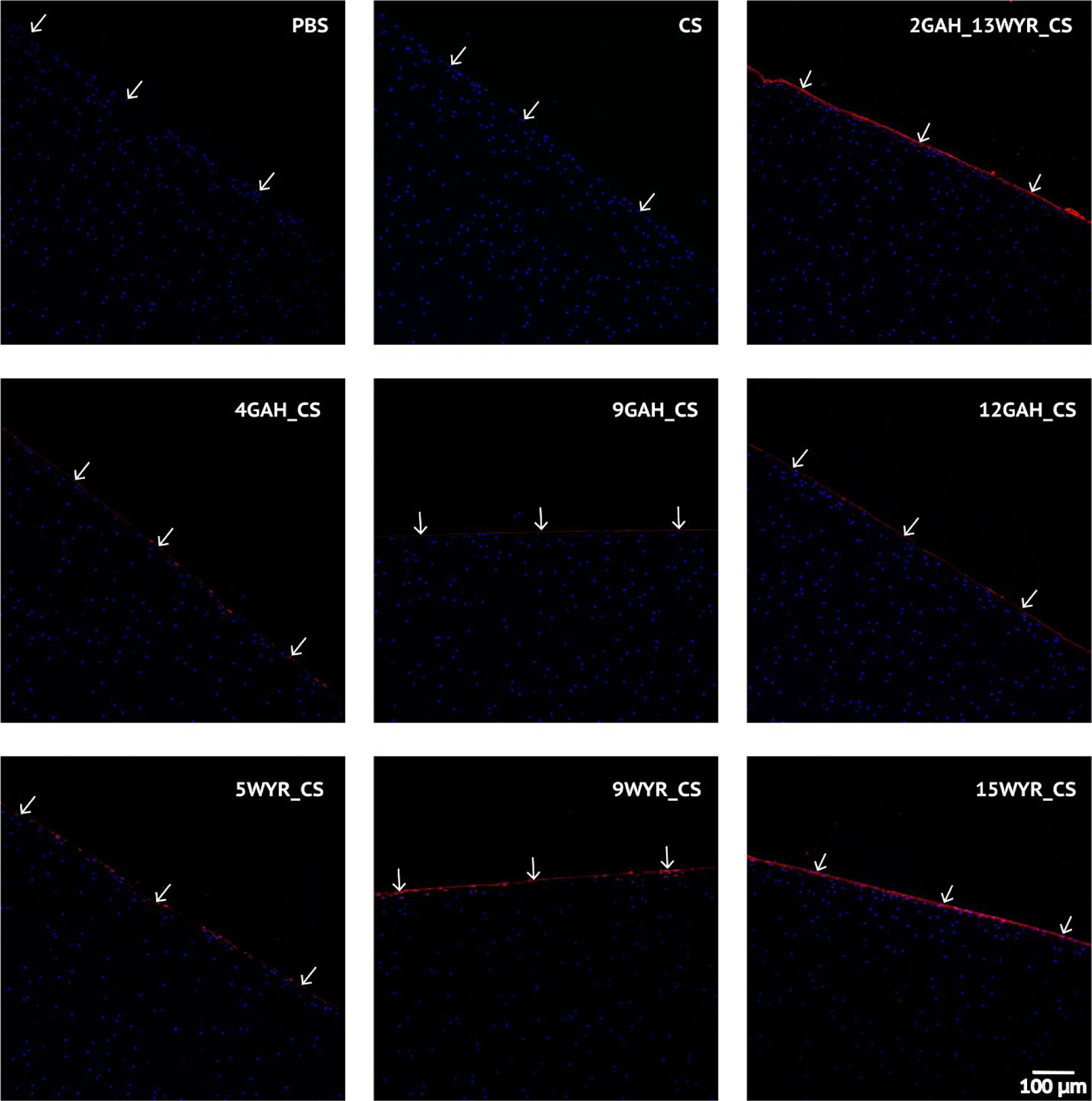
Figure 3.

Confocal imaging of post-rheology cartilage samples. The nuclei of chondrocytes were stained with DAPI (blue). The biotinylated peptidoglycan treatments at the cartilage surface were stained with Alexa Fluor® Streptavidin 633 (red). White arrows indicate cartilage surface.
