
Figure 5.
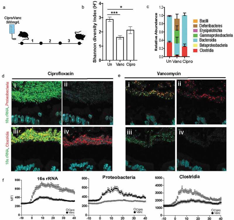
Dense community structure is sensitive to Vancomycin
A. Experimental design for comparing dense community structure in Ciprofloxacin and Vancomycin treated mice. Provided SPF WT mice with 500 mg/L Ciprofloxacin or Vancomycin in the drinking water for three days. B. Shannon diversity of untreated, Vancomycin, and Ciprofloxacin treated fecal communities (N = 6 mice per group, SD).C. Class-level relative abundance for untreated, Vancomycin, and Ciprofloxacin treated fecal communities (N = 6 mice per group, SEM).D. FISH images of spatial structure after Ciprofloxacin treatment showing preservation of community consisting of Clostridia and excluding Proteobacteria. i: merged image with host epithelium (DAPI, blue), all bacteria (16s rRNA, green), and Proteobacteria (red). ii. Merged image with host epithelium and Proteobacteria. iii. Merged image with host epithelium, all bacteria, and Clostridia (red). iv. Merged image with host epithelium and ClostridiaE. FISH images of spatial structure after Vancomycin treatment showing decimation of dense band of bacteria as well as the absence of Clostridia and the prevalence of Proteobacteria. i. merged image with host epithelium (DAPI, blue), all bacteria (16S rRNA, green), and Proteobacteria (red). ii: merged image with host epithelium and Proteobacteria. iii. Merged image with host epithelium, all bacteria, and Clostridia (red). iv. Merged image with host epithelium and ClostridiaF. Mean fluorescence intensity (MFI) measurements for representative mice quantifying bacterial density for all bacteria (16s rRNA), Proteobacteria, and Clostridia after Ciprofloxacin (open circles) and Vancomycin (closed circles) treatment. Measurements started 10-μm before start of bacterial signal closest to epithelial lining (N = 3 mice, SEM).