
FIGURE 1.
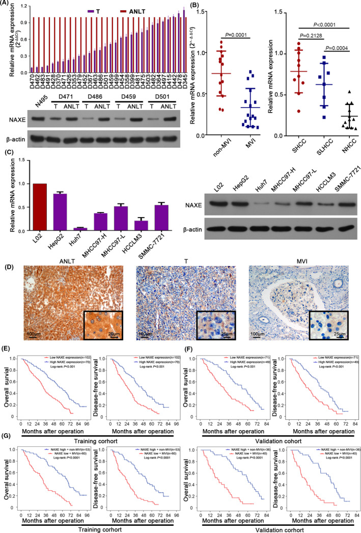
NAXE is significantly downregulated in HCC and is a promising prognostic marker for HCC. A, The mRNA and protein expression of NAXE in HCC tissues analyzed by qRT‐PCR and western blot. N495 indicates normal liver tissue. B, NAXE mRNA expression in patients with or without MVI and in SHCC, SLHCC and NHCC subgroups. C, The mRNA and protein expression of NAXE in L02 and HCC cell lines analyzed by qRT‐PCR and western blot. D, Representative immunohistochemistry images of NAXE in ANLT, HCC, and MVI. The black frames in the lower right corner show a higher magnification of the corresponding images. E and F, Kaplan‐Meier analysis (log‐rank test) for OS and DFS of HCC patients in training cohort (E) and validation cohort (F) according to NAXE expression level. G, Survival curves of HCC patients in training and validation cohort with high NAXE expression and absence of MVI or with low NAXE expression and presence of MVI. ANLT, adjacent nontumor liver tissue; MVI, microvascular invasion; NHCC, nodular HCC; SHCC, small HCC; SLHCC, solitary large HCC