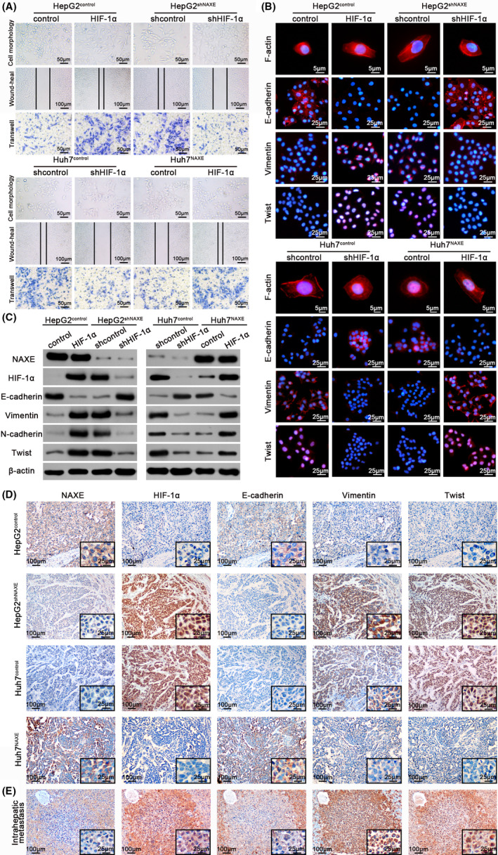
FIGURE 5.

Low NAXE expression promotes HCC migration, invasion, and EMT by activating HIF‐1α signaling. A, Representative images of cytomorphology, migration and invasion assay for NAXE‐interfered cells and control cells with HIF‐1α expression inhibited or restored. The data are quantified in Figure S7B,C. B, Fluorescence images of cytoskeleton, E‐cadherin, vimentin, and Twist for indicated cells with either HIF‐1α re‐inhibited or re‐introduced. Nuclei are identified by DAPI. C, The protein expression of EMT markers and Twist in indicated cells with either HIF‐1α re‐inhibited or re‐introduced. D and E, Representative immunohistochemistry images of indicated molecules using consecutive sections of liver orthotopic tumors (D) and intrahepatic metastatic nodule (E). The black frames in the lower right corner show higher magnification of corresponding images
